. 2014 Feb;21(2):143-51.
doi: 10.1038/nsmb.2740. Epub 2014 Jan 5.

The ABC-F protein EttA gates ribosome entry into the translation elongation cycle

Affiliations

The ABC-F protein EttA gates ribosome entry into the translation elongation cycle

Grégory Boël et al. Nat Struct Mol Biol. 2014 Feb.

Abstract

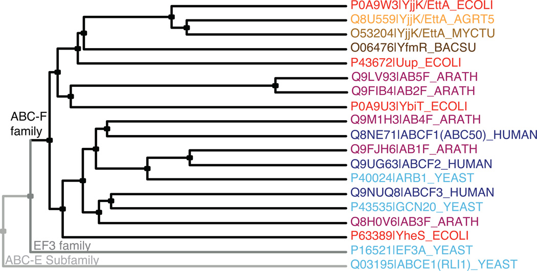
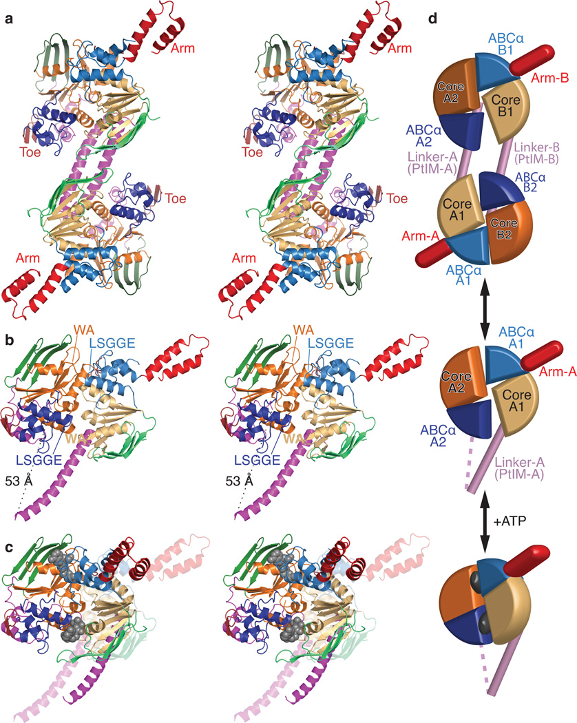
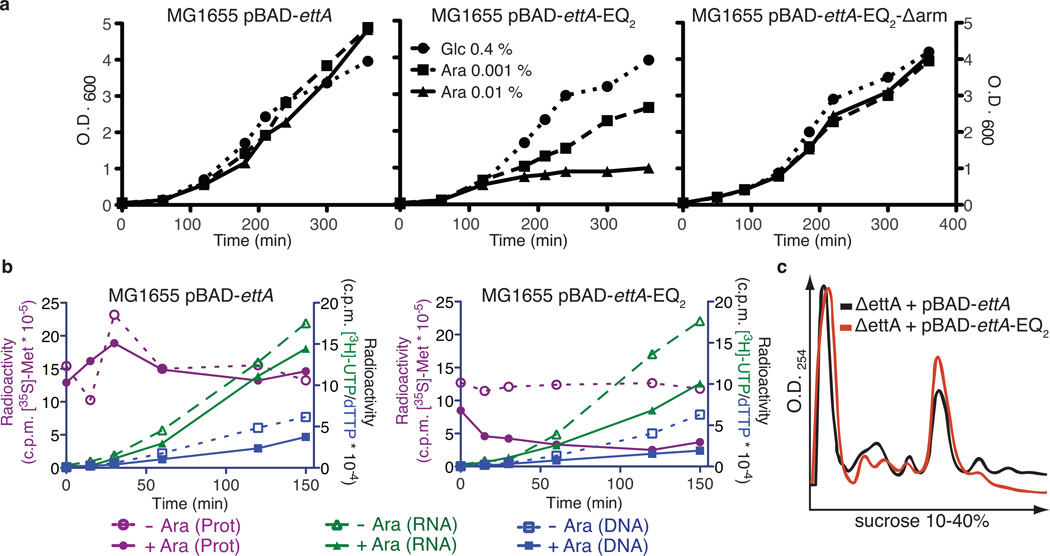
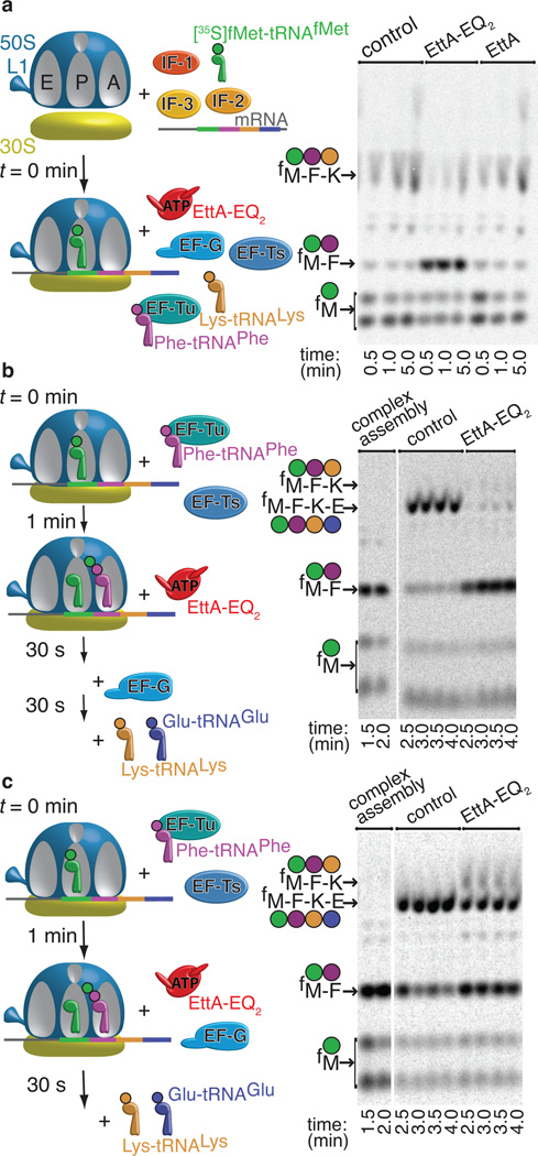
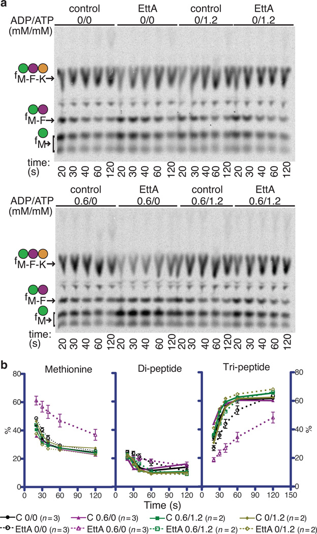
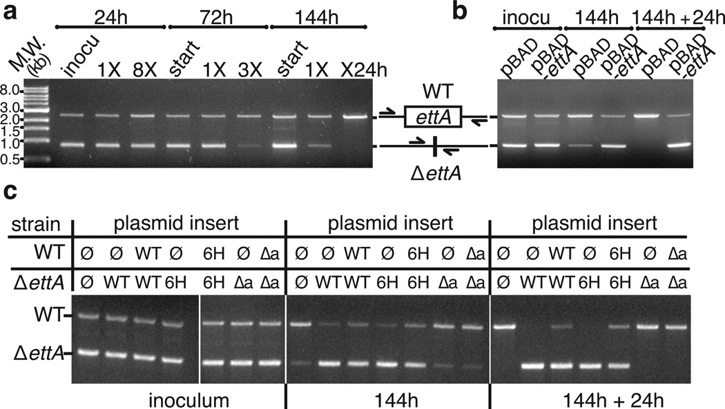
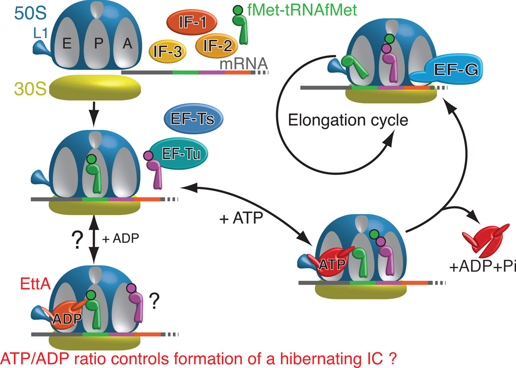
ABC-F proteins have evaded functional characterization even though they compose one of the most widely distributed branches of the ATP-binding cassette (ABC) superfamily. Herein, we demonstrate that YjjK, the most prevalent eubacterial ABC-F protein, gates ribosome entry into the translation elongation cycle through a nucleotide-dependent interaction sensitive to ATP/ADP ratio. Accordingly, we rename this protein energy-dependent translational throttle A (EttA). We determined the crystal structure of Escherichia coli EttA and used it to design mutants for biochemical studies including enzymological assays of the initial steps of protein synthesis. These studies suggest that EttA may regulate protein synthesis in energy-depleted cells, which have a low ATP/ADP ratio. Consistently with this inference, EttA-deleted cells exhibit a severe fitness defect in long-term stationary phase. These studies demonstrate that an ABC-F protein regulates protein synthesis via a new mechanism sensitive to cellular energy status.


Figures

Figure 1
Figure 1
ABC-F phylogeny. Cladogram produced using CLUSTAL-Ω and labeled with Swissprot species codes, which shows two bacterial orthologs of EttA (from A. tumefaciens and M. tuberculosis), one bacterial paralog of EttA (YfmR from B. subtillis), two non-ABC-F family proteins containing tandem ABC domains (eEF3 and ABCE1 from S. cerevisiae), and all ABC-F proteins from E. coli (red), S. cerevisiae (cyan), A. thaliana (purple), and H. sapiens (blue). Note that all of these ABC-F proteins, but neither eEF3 nor ABCE1, contain the PF12848 domain in addition to tandem ABC domains.
Figure 2
Figure 2
Crystal structure of E. coli EttA. (a) Stereopair showing the nucleotide-free EttA dimer in the asymmetric unit (Table 1). The ABC domains in each protomer are colored lighter (ABC1) and darker (ABC2) shades of similar colors (green for ABCβ, tan-orange for F1-like core, and blue for ABCα subdomains, red for the arm and toe motifs, and magenta for the PtIM). (b) Equivalently colored stereopair showing a magnified view of one interacting ABC1-ABC2 domain pair in the EttA dimer (generated by deleting 1-286 in protomer A and 278-555 in protomer B), which provides a model for the nucleotide-free conformation of the EttA monomer. Labels indicate the Walker A (WA) motif in the Fl-like core and the LSGGE signature sequence in the ABCα subdomain. The Walker B motif (Φ4DE, with Φ being any hydrophobe and terminating in catalytic base) is located between the WA and LSGGE motifs within each ABC. (c) Stereopair showing models for the nucleotide-free (translucent colors) and ATP-bound (solid colors) conformations of the EttA monomer superimposed via least-squares alignment of ABC2. The nucleotide-free conformation represents one ABC1-ABC2 domain pair from the crystallographically observed EttA dimer (panel b), while the ATP-bound conformation was modeled using rigid-body rotations to align the crystallographically observed nucleotide-free conformations of ABC1 and ABC2 to the two protomers in the ATP-sandwich dimer of the E171Q mutant of MJ0796; see “Structural Superposition” in Online Methods for details. (d) Schematics of the EttA dimer (top), nucleotide-free monomer (middle), and modeled ATP-bound monomer (bottom) colored as above.
Figure 3
Figure 3
Expression of EttA-EQ2 causes trans-dominant toxicity in vivo due to inhibition of protein synthesis. (a) Graphs showing OD600 profiles during expression of EttA variants in E. coli MG1655 in LB medium at 37 °C. Cells harboring pBAD-ettA, pBAD-ettA-EQ2, or pBAD-ettA-EQ2-Δarm plasmids were grown overnight in LB with 0.4% (w/v) glucose (Glc) to repress EttA expression and then diluted 1:100 into the same medium or alternatively one containing 0.001-0.01% (w/v) arabinose (Ara) to induce increasing levels of expression. (b) Graphs showing results from experiments using radiolabeled precursors to characterize the influence of expressing EttA variants on protein, RNA, and DNA synthesis in vivo. MG1655 cells harboring pBAD-ettA or pBAD-ettA-EQ2 plasmids were grown at 37 °C in M9 glycerol minimal medium to OD600 ~0.2 prior to induction of EttA expression using 0.2% (w/v) Ara at zero time on these graphs. RNA or DNA were labeled by adding [3H]UTP (green) or [3H]dTTP (blue), respectively, to the cultures at the same time as the inducer, while protein was labeled at the indicated time points by subjecting an aliquot of the culture to a 1 minute pulse with [35S]methionine (red). Cells were spotted onto a Whatman 3MM filter and washed with trichloroacetic acid (TCA) before scintillation counting of the radioactivity incorporated into polymers. (c) Plots of sucrose gradient profiles of polysomes from MG1655 ΔettA cells harboring pBAD-ettA or pBAD-ettA-EQ2 plasmids induced with 0.1% Ara for 30 minutes after reaching an OD600 of 0.6.
Figure 4
Figure 4
EttA-EQ2 inhibits translation after formation of the first peptide bound. Minimum in vitro translation assays were performed as explained in the schematics on the left at 37 °C in the presence of 0.5 mM ATP, 1.0 mM GTP, and a phophoenolpyruvate-based energy-regenerating system. Reaction products were analyzed by electrophoretic thin-layer chromatography (eTLC) and autoradiography (right). (a) After 70S IC formation, either buffer or 2.5 µM WT-EttA or EttA-EQ2 was added in parallel with the elongation factors, Phe-tRNAPhe and Lys-tRNALys. (b) After formation of the 70S IC and subsequent addition of EF-Tu, EF-Ts, and Phe-tRNAPhe to drive synthesis of the first peptide bond, either buffer or EttA-EQ2 was added 1 minute later, and the reaction was allowed to proceed for 30 seconds prior to the addition of EF-G, Lys-tRNALys, and Glu-tRNAGlu to enable tetrapeptide synthesis. (c) Same protocol as panel b, but to determine whether EF-G and EttA-EQ2 kinetically compete with one another, EF-G was added in parallel with buffer or EttA-EQ2 at 1 minute after addition of EF-Tu, EF-Ts, and Phe-tRNAPhe. Thirty seconds later, Lys-tRNALys and Glu-tRNAGlu were added to enable tetrapeptide synthesis. Reactions were conducted in Polymix Buffer (3.5 mM Mg(OAc)2, 100 mM KCl, 5 mM NH4OAc, 0.5 mM Ca(OAc)2, 0.1 mM EDTA, 1 mM spermidine, 5 mM putrescine, 6 mM 2-mercaptoethanol, 50 mM Tris-OAc, pH 6.9) using an mRNA template directing synthesis of an fMet-Phe-Lys-Glu (fMFKE) tetrapeptide.
Figure 5
Figure 5
WT-EttA inhibits synthesis of the first peptide bond at low ATP/ADP ratio. (a) Room temperature in vitro translations with or without 0.6 mM ADP and 1.2 mM ATP were analyzed by eTLC. Reactions, conducted as in Fig. 4a but with the 70S IC desalted in Polymix Buffer, contained 0.3 mM GTP, 0.6 µM 70S ribosomes, and when indicated 3.5 µM of an EttA variant added in parallel with the elongation factors, Phe-tRNAPhe, and Lys-tRNALys. (b) Quantification of products in the autoradiograms in panel a using ImageQuant software, with error bars representing standard error of the mean.
Figure 6
Figure 6
WT and His6-EttA promote survival in long-term stationary phase. Agarose gels are shown that visualize PCR products quantifying the relative population of wild-type vs. ΔettA cells in competitive fitness assays in LB at 37 °C. The chromosomal region flanking ettA by 400 basepairs was amplified from total DNA in mixed cultures. (a) Starting cultures containing a 1:1 mixture of overnights from the individual strains were grown for 24, 72, or 144 hours prior to re-inoculation into fresh medium and re-growth for the same period of time. Eight growth cycles were performed for the 24-hour culture, three for the 72-hour culture, and two for the 144-hour culture, which was re-grown for an additional 24 hours prior to analysis. (b) Mixed cultures of the ΔettA strain containing pBAD or pBAD-ettA plasmids were grown for 144 hours, prior to re-inoculation for an additional 24 hours. Immunoblotting analysis with anti-EttA antibody (unpublished) demonstrates a roughly physiological level of expression from the pBAD-ettA plasmid under these growth conditions (i.e., without supplementation with glucose to repress expression from the arabinose promoter controlling expression of EttA). (c) Results from equivalent complementation experiments performed on mixed cultures of the ΔettA and wild-type strains containing pBAD plasmid with different inserts (ø: no insert, WT: WT-EttA, 6H: His6-EttA, Δa: EttA-Δarm).
Figure 7
Figure 7
Schematic model of EttA function based on the results presented here and in the companion paper. In the presence of ADP, EttA inhibits formation of the first peptide bound (Fig. 5b), which may be mediated by stabilization of the 70S IC in a hibernating state by ADP-bound EttA. (See Discussion). In contrast, ATP-bound EttA stimulates the formation of the first peptide bond by the ribosome and then, concomitantly with ATP hydrolysis, dissociates from the ribosome, thereby allowing it to enter the elongation cycle.

Comment in

  • The ABCs of the ribosome.
    Fredrick K, Ibba M. Fredrick K, et al. Nat Struct Mol Biol. 2014 Feb;21(2):115-6. doi: 10.1038/nsmb.2765. Nat Struct Mol Biol. 2014. PMID: 24500425 Free PMC article. No abstract available.

References

    1. Davidson AL, Dassa E, Orelle C, Chen J. Structure, function, and evolution of bacterial ATP-binding cassette systems. Microbiol Mol Biol Rev. 2008;72:317–364. table of contents. - PMC - PubMed
    1. Cavanaugh LF, Palmer AG, 3rd, Gierasch LM, Hunt JF. Disorder breathes life into a DEAD motor. Nat Struct Mol Biol. 2006;13:566–569. - PubMed
    1. Jones PM, George AM. Subunit interactions in ABC transporters: towards a functional architecture. FEMS Microbiol Lett. 1999;179:187–202. - PubMed
    1. Hopfner KP, et al. Structural biology of Rad50 ATPase: ATP-driven conformational control in DNA double-strand break repair and the ABC-ATPase superfamily. Cell. 2000;101:789–800. - PubMed
    1. Smith PC, et al. ATP binding to the motor domain from an ABC transporter drives formation of a nucleotide sandwich dimer. Mol Cell. 2002;10:139–149. - PMC - PubMed

Publication types

MeSH terms

Substances

Associated data