. 2014 Mar 4;21(2):297-310.
doi: 10.1530/ERC-13-0339. Print 2014 Apr.

YAP regulates cell proliferation, migration, and steroidogenesis in adult granulosa cell tumors

Affiliations

YAP regulates cell proliferation, migration, and steroidogenesis in adult granulosa cell tumors

David Fu et al. Endocr Relat Cancer. .

Abstract

The Hippo signaling pathway has been implicated as a conserved regulator of organ size in both Drosophila and mammals. Yes-associated protein (YAP), the central component of the Hippo signaling cascade, functions as an oncogene in several malignancies. Ovarian granulosa cell tumors (GCT) are characterized by enlargement of the ovary, excess production of estrogen, a high frequency of recurrence, and the potential for malignancy and metastasis. Whether the Hippo pathway plays a role in the pathogenesis of GCT is unknown. This study was conducted to examine the expression of YAP in human adult GCTs and to determine the role of YAP in the proliferation and steroidogenesis of GCT cells. Compared with age-matched normal human ovaries, GCT tissues exhibited higher levels of YAP expression. YAP protein was predominantly expressed in the nucleus of tumor cells, whereas the non-tumor ovarian stromal cells expressed very low levels of YAP. YAP was also expressed in cultured primary human granulosa cells and in KGN and COV434 GCT cell lines. siRNA-mediated knockdown of YAP in KGN cells resulted in a significant reduction in cell proliferation (P<0.001). Conversely, overexpression of wild type YAP or a constitutively active YAP (YAP1) mutant resulted in a significant increase in KGN cell proliferation and migration. Moreover, YAP knockdown reduced FSH-induced aromatase (CYP19A1) protein expression and estrogen production in KGN cells. These results demonstrate that YAP plays an important role in the regulation of GCT cell proliferation, migration, and steroidogenesis. Targeting the Hippo/YAP pathway may provide a novel therapeutic approach for GCT.

Keywords: Hippo/YAP pathway; cell proliferation; granulosa cell tumor; migration; steroidogenesis.


Figures

Figure 1
Figure 1
YAP is expressed in human granulosa cell tumors. Immunohistochemistry was used to examine the expression of YAP in normal ovarian tissues (A, C, E) and human granulosa cell tumor tissues (B & D, F). YAP staining is indicated in brown. The counter stain is hematoxylin shown in blue. A) Whole tissue section scan of a representative normal ovary stained with YAP antibody; B) Scanned image of partial ovarian granulosa cell tumor tissue stained with YAP. Amplified representative images of normal ovarian tissues (C) and GCT tissues (E) stained with YAP antibody to show cell-type specific YAP expression. Representative high-resolution images were also presented to show the subcellular localization of YAP in the normal ovarian tissues (E) and GCT tissues (F). Tissue sections probed with the same amount of non-immunized rabbit IgG was used as antibody control (G). Quantitative results of DAB intensity were also presented to show the difference of positivity (percentage of the number of DAB positive cells relative to number of total cells in each tissue section) and relative intensity of YAP immunosignal between normal human ovarian tissues (CTRL) and tumor tissues (GCT). Each bar represents the mean ± SEM. *: significantly different (P < 0.001) from the control (CTRL) groups. Scale bar = 2 mm in A & B; scale bar = 100 μm in C & D; scale bar=50 μm in E & F; SC: stromal cells; CL: corpus luteum cells, TC: tumor cells.
Figure 2
Figure 2
YAP is expressed in human ovarian GCT cell lines and epithelial ovarian cancer cell lines. A) Localization of YAP protein in KGN cells using fluorescent immunocytochemistry. A1) Total YAP (green); A2) YAP protein (green) was merged with actin filaments (red, stained with Rhodamine-phalloidin). The nucleus was stained with DAPI (blue). A3) KGN cells stained with same amount of rabbit IgG (antibody control) and Rhodamine-phalloidin. Scale bar = 25 μm. B) RT-PCR detection of YAP mRNA expression in cultured human granulosa cells (human-GC), GCT cell lines (KGN and COV434) and ovarian cancer cell lines (IGROV1, SKOV-3 and CAOV-3). C) Western blot detection of YAP protein expression in the same cell lines. p-YAP: Serine127 phosphorylated YAP protein. GAPDH and tubulin were used as mRNA or protein loading control, respectively.
Figure 3
Figure 3
YAP is required for KGN cell proliferation. A) Fluorescence-labeled scramble siRNA (siGLO) indicates siRNA transfection efficiency in KGN cells. B) Western blot shows the down regulation of YAP protein after YAP siRNA treatments. β-tubulin was used as a protein loading control. The lower panel is the quantitative results of Western blot. Bars represent means ± SEM (n = 3). C) Morphology of siGLO transfected KGN cells and YAP siRNA transfected cells. Scale bar = 100 μm. D) KGN cells were transfected with YAP siRNAs for 6 hours and then cultured for another 72 hours. Cell numbers in each group were determined with an INVITROGEN Countess ® cell counter. Each bar represents the mean ± SEM (n = 5). Bars with same letter are not significantly (P > 0.05) different from each other.
Figure 4
Figure 4
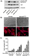
Overexpression of YAP stimulates KGN cell proliferation. A) Empty plasmid transfected KGN cells (KGN-MX), wild-type YAP overexpression cells (KGN-YAP) and mutant YAP transfected KGN cells (KGN-YAP-S127A) were plated in 6 well plates and the levels of YAP and phosphorylated YAP in these established stable cell lines were examined by Western blot. β-tubulin was used as a protein loading control. B) Upper panel: morphological difference between mutant YAP transfected KGN cells (KGN-YAP-S127A) and Empty plasmid transfected KGN cells (KGN-MX). Scale bar = 100 μm. Lower panel: actin filaments stained with rhodamine-phalloidin to demonstrate the morphological change between two established cell lines. Nuclei were stained with DAPI. Scale bar = 25 μm. C) Effect of YAP overexpression on KGN cell proliferation. Wild type KGN cells (KGN-MX), KGN-YAP cells and YAP-S127A-KGN cells were incubated for 24, 48 and 72 h and the cell number was counted with an INVITROGEN Countess ® automatic cell counter. Each bar represents the mean ± SEM, n = 5. Bars with same letter are not significantly (P > 0.05) different from each other.
Figure 5
Figure 5
YAP regulation of KGN cell migration. A) KGN-MXIV cells, KGN-YAP cells and KGN-YAPS127A cells were loaded to a transwell migration assay chamber and the migration assay was performed as described in the Materials and Methods section. Top panel, representative image of the migrated KGN-MXIV cells. Lower panel, representative image of the migrated KGN-YAPS127A cells. Scale bar, 100 μm. B) Migrated cells in each group were counted and the fold change (relative to the KGN-MXIV control group) of the migrated cells was determined. Each bar represents the mean ± SEM (n =12). Bars with same letter are not significantly (P > 0.05) different from each other. C) Evaluation of the cell migration ability with the wound-healing assay. Representative images from triplicate experiments were presented. Scale bar, 100μm
Figure 6
Figure 6
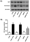
YAP is required for FSH-stimulated estradiol secretion. Normal KGN cells (CTRL), siGLO-transfected KGN cells and YAP siRNA transfected KGN cells were incubated with or without 100 ng/ml of FSH for 48 h. The protein level of YAP and aromatase were determined by western blot. β-tubulin was used as a protein loading control. 17β-estradiol in the medium was measured by ELISA. Each bar represents the mean ± SEM, n = 5. Bars with same letter are not significantly (p>0.05) different from each other. CTRL: Normal KGN cell as control. E2: 17β-estradiol.

References

    1. Aboud E. A review of granulosa cell tumours and thecomas of the ovary. Arch Gynecol Obstet. 1997;259:161–165. - PubMed
    1. Adashi EY, Hsueh AJ. Estrogens augment the stimulation of ovarian aromatase activity by follicle-stimulating hormone in cultured rat granulosa cells. J Biol Chem. 1982;257:6077–6083. - PubMed
    1. Al-Agha OM, Huwait HF, Chow C, Yang W, Senz J, Kalloger SE, Huntsman DG, Young RH, Gilks CB. FOXL2 is a sensitive and specific marker for sex cord-stromal tumors of the ovary. Am J Surg Pathol. 2011;35:484–494. - PubMed
    1. Alhilli MM, Long HJ, Podratz KC, Bakkum-Gamez JN. Aromatase inhibitors in the treatment of recurrent ovarian granulosa cell tumors: brief report and review of the literature. J Obstet Gyaecol Res. 2012;38:340–344. - PubMed
    1. Amsterdam A, Selvaraj N. Control of differentiation, transformation, and apoptosis in granulosa cells by oncogenes, oncoviruses, and tumor suppressor genes. Endocr Rev. 1997;18:435–461. - PubMed

Publication types

MeSH terms

Supplementary concepts