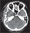
The supposed intracavernous sinus arachnoid cyst with abducens neuropathy: a case report
- PMID: 24390178
- PMCID: PMC4533470
- DOI: 10.2176/nmc.cr.2013-0092
The supposed intracavernous sinus arachnoid cyst with abducens neuropathy: a case report
Abstract
Intracavernous sinus arachnoid cysts are rare intracranial congenital lesions. When present, their anatomic location frequently results in cranial nerve palsy. A 15-year-old boy was admitted to our hospital with diplopia, which had gradually worsened over the previous several months. An arachnoid cyst was identified within the right cavernous sinus and fenestration surgery was performed. The patient recovered well and three months after the surgery, diplopia was disappeared. Surgical decompression of the intracavernous sinus arachnoid cyst is beneficial for symptomatic patients with this condition.
Conflict of interest statement
The authors have no personal, financial, or institutional interest in any of the drugs, materials, or devices in the article. All authors who are members of The Japan Neurosurgical Society (JNS) have registered online Self-reported COI Disclosure Statement Forms through the website for JNS members.
Figures


Similar articles
-
Progressive sixth nerve palsy secondary to intracavernous arachnoid cyst and complicated by contralateral optic nerve sheath meningioma.Eur J Ophthalmol. 2020 Sep;30(5):NP86-NP89. doi: 10.1177/1120672119853133. Epub 2019 Jun 3. Eur J Ophthalmol. 2020. PMID: 31155935
-
Cranial Nerve Palsies in the Setting of Arachnoid Cysts: A Case Series and Literature Review.J Neuroophthalmol. 2024 Jun 1;44(2):242-246. doi: 10.1097/WNO.0000000000001983. Epub 2023 Sep 1. J Neuroophthalmol. 2024. PMID: 37656595 Review.
-
Intracavernous sinus arachnoid cyst with optic neuropathy.J Clin Neurosci. 2010 Feb;17(2):267-9. doi: 10.1016/j.jocn.2009.05.027. Epub 2009 Dec 29. J Clin Neurosci. 2010. PMID: 20036550
-
Sixth cranial nerve palsy due to arachnoid cyst.J Pediatr Ophthalmol Strabismus. 2014 Oct 1;51 Online:e58-61. doi: 10.3928/01913913-20140923-02. J Pediatr Ophthalmol Strabismus. 2014. PMID: 25347081
-
Intracavernous schwannoma of the abducens nerve: a review of the clinical features, radiology and pathology of an unusual case.J Clin Neurosci. 2001 Jul;8(4):357-60. doi: 10.1054/jocn.2000.0846. J Clin Neurosci. 2001. PMID: 11437580 Review.
Cited by
-
Pupil-sparing third cranial nerve palsy with aberrant regeneration secondary to cavernous sinus arachnoid cyst.eNeurologicalSci. 2018 Nov 19;14:28-30. doi: 10.1016/j.ensci.2018.11.017. eCollection 2019 Mar. eNeurologicalSci. 2018. PMID: 30555949 Free PMC article.
References
-
- Leo JS, Pinto RS, Hulvat GF, Epstein F, Kricheff II: Computed tomography of arachnoid cysts. Radiology 130: 675– 680, 1979. - PubMed
-
- Duz B, Kaya S, Daneyemez M, Gonul E: Surgical management strategies of intracranial arachnoid cysts: a single institution experience of 75 cases. Turk Neurosurg 22: 591– 598, 2012. - PubMed
-
- Barr D, Kupersmith MJ, Pinto R, Turbin R: Arachnoid cyst of the cavernous sinus resulting in third nerve palsy. J Neuroophthalmol 19: 249– 251, 1999. - PubMed
-
- Cheng CH, Lin HL, Cho DY, Chen CC, Liu YF, Chiou SM: Intracavernous sinus arachnoid cyst with optic neuropathy. J Clin Neurosci 17: 267– 269, 2010. - PubMed
-
- Kobayashi T, Negoro M, Awaya S: Cavernous sinus subarachnoid diverticulum and sixth nerve palsy. Neuroradiology 29: 306– 307, 1987. - PubMed
Publication types
MeSH terms
LinkOut - more resources
Full Text Sources
Other Literature Sources

