. 2014 Mar;88(6):3273-84.
doi: 10.1128/JVI.03478-13. Epub 2014 Jan 3.

Viral entry of hepatitis B and D viruses and bile salts transportation share common molecular determinants on sodium taurocholate cotransporting polypeptide

Affiliations

Viral entry of hepatitis B and D viruses and bile salts transportation share common molecular determinants on sodium taurocholate cotransporting polypeptide

Huan Yan et al. J Virol. 2014 Mar.

Abstract

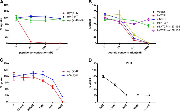
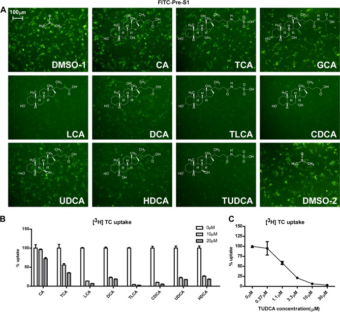
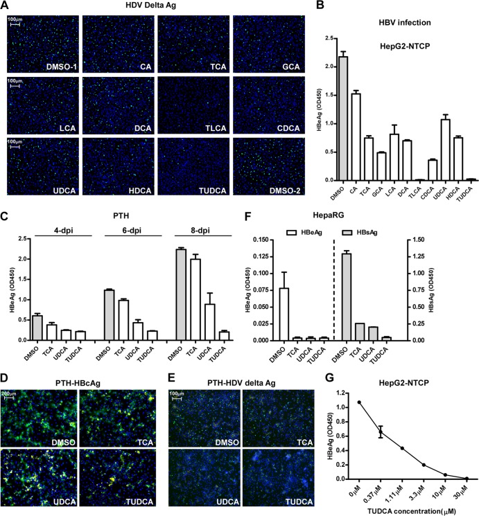
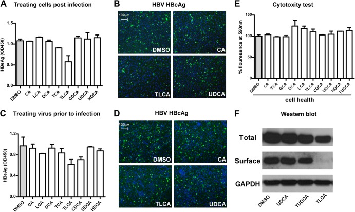
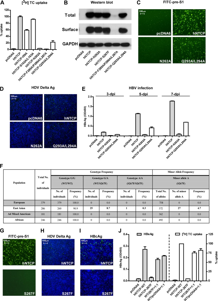
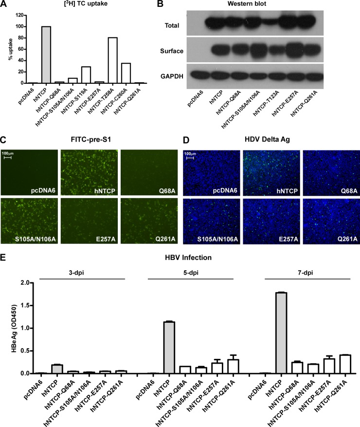
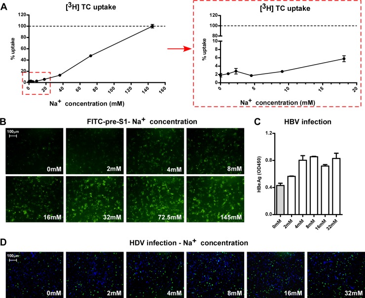
The liver bile acids transporter sodium taurocholate cotransporting polypeptide (NTCP) is responsible for the majority of sodium-dependent bile salts uptake by hepatocytes. NTCP also functions as a cellular receptor for viral entry of hepatitis B virus (HBV) and hepatitis D virus (HDV) through a specific interaction between NTCP and the pre-S1 domain of HBV large envelope protein. However, it remains unknown if these two functions of NTCP are independent or if they interfere with each other. Here we show that binding of the pre-S1 domain to human NTCP blocks taurocholate uptake by the receptor; conversely, some bile acid substrates of NTCP inhibit HBV and HDV entry. Mutations of NTCP residues critical for bile salts binding severely impair viral infection by HDV and HBV; to a lesser extent, the residues important for sodium binding also inhibit viral infection. The mutation S267F, corresponding to a single nucleotide polymorphism (SNP) found in about 9% of the East Asian population, renders NTCP without either taurocholate transporting activity or the ability to support HBV or HDV infection in cell culture. These results demonstrate that molecular determinants critical for HBV and HDV entry overlap with that for bile salts uptake by NTCP, indicating that viral infection may interfere with the normal function of NTCP, and bile acids and their derivatives hold the potential for further development into antiviral drugs.

Importance: Human hepatitis B virus (HBV) and its satellite virus, hepatitis D virus (HDV), are important human pathogens. Available therapeutics against HBV are limited, and there is no drug that is clinically available for HDV infection. A liver bile acids transporter (sodium taurocholate cotransporting polypeptide [NTCP]) critical for maintaining homeostasis of bile acids serves as a functional receptor for HBV and HDV. We report here that the NTCP-binding lipopeptide that originates from the first 47 amino acids of the pre-S1 domain of the HBV L protein blocks taurocholate transport. Some bile salts dose dependently inhibit HBV and HDV infection mediated by NTCP; molecular determinants of NTCP critical for HBV and HDV entry overlap with that for bile acids transport. This work advances our understanding of NTCP-mediated HBV and HDV infection in relation to NTCP's physiological function. Our results also suggest that bile acids or their derivatives hold potential for development into novel drugs against HBV and HDV infection.


Figures

FIG 1
FIG 1
Binding of the HBV pre-S1 domain to NTCP impairs taurocholate uptake. (A) Inhibition of [3H]taurocholate uptake by HBV pre-S1 lipopeptide. A [3H]taurocholate uptake assay was conducted with HepG2-hNTCP cells (HepG2 cells stably expressing human NTCP) pretreated with the indicated concentrations of HBV pre-S1 lipopeptide or its derivatives at 37°C for 2 h. myr(+)47, myristoylated pre-S1 peptide containing the first N-terminal 47 amino acids of the L protein of HBV; myr(−)47, the pre-S1 peptide without myristoylation modification; myr(+)47-N9K, myristoylated pre-S1 peptide with an asparagine (N)-to-arginine (K) mutation at residue 9. Cellular uptake of [3H]taurocholate in the absence of any peptide was set to 100%. (B) Inhibition of [3H]taurocholate uptake by myr(+)47 in HepG2 cells transiently transfected with NTCPs of different species. Cellular uptake of [3H]taurocholate by human NTCP when treated with dimethyl sulfoxide (DMSO) was set to 100%. (C) Inhibition of [3H]taurocholate uptake by cholesterol or myristoyl-modified pre-S1 peptides in HepG2-NTCP cells. Uptake of [3H]taurocholate by cells treated with DMSO was set to 100%. (D) Inhibition of [3H]taurocholate uptake by myristoylated pre-S1 peptide in primary Tupaia hepatocytes.
FIG 2
FIG 2
Bile salts blocked the interaction between the pre-S1 peptide and NTCP. (A) Inhibition of FITC-conjugated pre-S1 peptide binding by different bile salts. FITC–pre-S1 peptide (400 nM) was incubated with NTCP-transfected HepG2 cells for 1 h in the presence of the indicated bile salt (20 μM). The binding efficiency of the pre-S1 peptide was analyzed via a fluorescence microscope. CA, cholic acid; GCA, glycocholic acid; LCA, lithocholic acid; DCA, deoxycholic acid; TCA, taurocholic acid; TLCA, taurolithocholic acid; CDCA, chenodeoxycholic acid; UDCA, ursodeoxycholic acid; HDCA, hyodeoxycholic acid; TUDCA, tauroursodeoxycholic acid. (B) Competition for [3H]taurocholate uptake by other bile salts. The indicated bile salts (at 10 μM or 20 μM) were examined. Uptake efficiency is presented as the percent uptake compared with the dimethyl sulfoxide group. (C) The ability of TUDCA to inhibit [3H]taurocholate uptake by HepG2-NTCP cells. TUDCA at the indicated concentration was present in the medium during the entire [3H]taurocholate uptake process.
FIG 3
FIG 3
Bile acids inhibited HBV and HDV infection. (A and B) Inhibition of HDV (A) and HBV (B) infection in HepG2-NTCP cells in the presence of bile salts. HepG2-NTCP cells were infected with HDV at 37°C for 24 h in the presence of 5% PEG 8000 and 20 μM of each of the indicated bile salts except for LCA (tested at 5 μM). The intracellular HDV delta antigen was stained with mouse monoclonal antibody 4G5 at day 8 p.i. The level of secreted HBeAg in the culture medium collected at day 7 p.i. was measured with an ELISA kit. (C to E) Inhibition of HDV and HBV infection in primary Tupaia hepatocytes by 20 μM TCA, UDCA, or TUDCA. PTHs were infected with HDV or HBV in the presence of 20 μM bile salt. For HBV infection, the level of secreted HBeAg in the culture medium was evaluated at 3, 5, and 7 days p.i. (C). On day 8 p.i., HBV core antigen of the infected cells were stained with MAb 1C10 (green) (D). For HDV, intracellular HDV delta antigen of the infected cells at day 8 p.i. were stained with MAb 4G5 (green) (E). The yellow fluorescence in panels D and E stands for the autofluorescence signal from dead cells, which existed in both the green and red channels. (F) Inhibition of HBV infection of human hepatoma HepaRG cells in the presence of 25 μM bile salts. The levels of HBeAg and HBsAg in the culture medium at 6 days p.i. were measured via an ELISA. (G) Dose-dependent inhibition of HBV infection by TUDCA in HepG2-NTCP cells. The secreted HBeAg at 6 days p.i. was measured in an ELISA.
FIG 4
FIG 4
Analysis of the direct effects of bile salts on HepG2-NTCP cells and HBV. (A and B) HBV infection of HepG2-NTCP cells that were treated with bile salts post-HBV inoculation. HepG2-NTCP cells were inoculated with HBV at 37°C for 24 h in the presence of 5% PEG 8000. After the inoculation, cells were washed and then incubated with different bile salts at the indicated concentrations for another 24 h, and then culturing of cells was continued in PMM. HBeAg in the culture medium collected at 7 days p.i. was measured in an ELISA (A). On day 8 p.i., HBcAg in the infected cells treated with the indicated bile salts was stained with anticore MAb 1C10 (green), and the nuclei were stained with DAPI (blue) (B). (C and D) Infection of HepG2-NTCP cells treated with bile salts prior to HBV inoculation. The viruses were incubated with 20 μM bile salts for 24 h, then precipitated with 8% PEG 8000 to remove the bile salts, followed by resuspending in PMM. The treated viruses were then used to inoculate HepG2-NTCP cells for infection. The secreted HBeAg in the culture medium collected at 7 days p.i. was measured in an ELISA (C). On day 8 p.i., HBcAg in the infected cells was stained green with 1C10 MAb, and the nuclei were stained with DAPI (blue) (D). (E) Cytotoxicy analysis of the bile salts for HepG2-NTCP cells. HepG2-NTCP cells were treated with 20 μM bile salts (5 μM for LCA) in PMM for 24 h in the presence of 5% PEG 8000. Cells were then incubated with 10% alamarBlue (Invitrogen) in PMM for 1 h. Cellular health was evaluated based on fluorescence absorbance at 590 nm. The absorbance of dimethyl sulfoxide-treated cells was set to 100%. (F) Total and surface expression levels of NTCP from HepG2-NTCP cells in response to the indicated bile salt treatments.
FIG 5
FIG 5
NTCP mutants, corresponding to the residues important for bile salts binding/uptake, inhibited viral infections by HDV and HBV. (A) [3H]taurocholate uptake efficiency of HepG2 cells complemented with various NTCP mutants. HepG2 cells were transfected with NTCP mutants, followed by further culturing in PMM for 24 h before examination of their [3H]taurocholate uptake efficiency. (B) Total and surface expression levels of NTCP mutants in HepG2 cells. HepG2 cells were transfected with the indicated NTCP variants and then cultured in PMM for 24 h. Cells were then lysed by RIPA buffer, and the total and surface expression levels were analyzed by Western blotting. The expression level of GAPDH was used as an internal control. (C) FITC–pre-S1 binding efficiency of NTCP variants. HepG2 cells transfected as described for panel A were incubated with 400 nM FITC–pre-S1 at 37°C for 2 h, and the images were captured with a fluorescence microscope. (D) HDV infection efficiency for HepG2 cells transfected with NTCP variants. The intracellular delta antigen was stained with MAb 4G5 at 8 days p.i. (E) HBV infection of HepG2 cells expressing NTCP variants. The levels of HBeAg in the culture medium from HBV-infected HepG2 cells expressing the indicated NTCP variants were measured at 3, 5, and 7 days p.i. (F) Genotype frequency and allele frequency of the SNP that causes the NTCP S267F variant in different populations; the variant is caused by a single-nucleotide G-to-A change (rs2296651). The SNP is located at chromosome 14:70245193 (Ensembl Homo sapiens version 73.37) and corresponds to a C-to-T change in codon 267 (from TCC to TTC) on the reverse strand encoding human NTCP. According to data from the 1000 Genomes Project (http://www.1000genomes.org), in East Asia the heterozygous genotype frequency is 8.7%, and the minor allele homozygous genotype frequency is 0.3%. No SNP at this position is observed in other populations. The genotype distribution in East Asian population conforms to a Hardy-Weinberg equilibrium, which was verified by a chi-square test (P = 0.22). WT, wild type; Ad mixed, populations with recent ancestry from two or more genetically separated populations. (G) FITC–pre-S1 binding efficiency of NTCP in cells complemented with wild-type NTCP or a NTCP-S267F variant. (H and I) HDV and HBV infection efficiencies on HepG2 cells transfected with NTCP or NTCP-S267F. The intracellular delta antigen in the infected cells was stained with MAb 4G5 at 8 days p.i. (H). HBcAg was evaluated based on intracellular HBcAg at 8 days p.i. (I). (J) HBV infection efficiency and [3H]taurocholate uptake efficiency of HepG2 cells transfected with plasmid carrying wild-type (WT) NTCP, NTCP variant S267F, pcDNA6, or a mixture of the wild-type NTCP with either the S267F variant or pcDNA6 plasmid at a ratio of 1:1. The total amount of DNA for all the transfections was 1 μg DNA for ∼1.7 × 10⁵ HepG2 cells. The level of secreted HBeAg at 6 days p.i. was measured in an ELISA (left), and the [3H]taurocholate uptake efficiency at 24 h posttransfection was determined by liquid scintillation (wild-type NTCP was set to 100%) (right).
FIG 6
FIG 6
NTCP mutants corresponding to the residues important for sodium binding inhibit viral infections by HDV and HBV. (A) [3H]taurocholate uptake efficiency of HepG2 cells complemented with NTCP variants. (B) Western blot analysis of total and surface expression levels of the NTCP variants in HepG2 cells. The expression level of GAPDH was used as an internal control. (C) FITC–pre-S1–binding efficiency in HepG2 cells expressing NTCP variants. (D and E) HDV and HBV infection efficiencies in HepG2 cells expressing the NTCP variants. The intracellular HDV delta antigen in infected cells was stained by 4G5 at 8 days p.i. (D). Secreted HBeAg in the culture medium at 3, 5, and 7 days p.i. was measured in an ELISA (E).
FIG 7
FIG 7
Effects of extracellular Na+ concentration on the binding of pre-S1 to NTCP and viral infections. (A) Uptake of [3H]taurocholate in Ringer's solution containing increasing concentrations of Na+. The [3H]taurocholate uptake assay was conducted with HepG2-NTCP cells in Ringer's solution for 15 min (left). The level of uptake at 145 mM was set to 100%. (Right) Uptake efficiency in response to Na+ concentrations lower than 20 mM. (B) FITC–pre-S1 peptide-binding efficiency in different Na+ concentrations. HepG2 cells transfected with NTCP were incubated with 400 nM FITC–pre-S1 peptide in Ringer's solution containing different concentrations of Na+ for 10 min. Images were taken using a fluorescence microscope after extensive washing. (C and D) Na+ binding is required for efficient HBV and HDV infection. HBV and HDV were precipitated by 8% PEG 8000, resuspended in choline+ Ringer's solution, and then the Na+ concentration was adjusted as indicated. The viruses were then used to inoculate HepG2-NTCP cells for 6 h. HBV infection was assessed based on secreted HBeAg at 6 days p.i. in an ELISA (C). HDV infection was visualized by staining of the intracellular delta antigen at 8 days p.i. (D).

References

    1. Ott JJ, Stevens GA, Groeger J, Wiersma ST. 2012. Global epidemiology of hepatitis B virus infection: new estimates of age-specific HBsAg seroprevalence and endemicity. Vaccine 30:2212–2219. 10.1016/j.vaccine.2011.12.116 - DOI - PubMed
    1. Hughes SA, Wedemeyer H, Harrison PM. 2011. Hepatitis delta virus. Lancet 378:73–85. 10.1016/S0140-6736(10)61931-9 - DOI - PubMed
    1. Yan H, Zhong G, Xu G, He W, Jing Z, Gao Z, Huang Y, Qi Y, Peng B, Wang H, Fu L, Song M, Chen P, Gao W, Ren B, Sun Y, Cai T, Feng X, Sui J, Li W. 2012. Sodium taurocholate cotransporting polypeptide is a functional receptor for human hepatitis B and D virus. eLife 1:e00049. 10.7554/eLife.00049 - DOI - PMC - PubMed
    1. Zhong G, Yan H, Wang H, He W, Jing Z, Qi Y, Fu L, Gao Z, Huang Y, Xu G, Feng X, Sui J, Li W. 2013. Sodium taurocholate cotransporting polypeptide mediates woolly monkey hepatitis B virus infection of Tupaia hepatocytes. J. Virol. 87:7176–7184. 10.1128/JVI.03533-12 - DOI - PMC - PubMed
    1. Yan H, Peng B, He W, Zhong G, Qi Y, Ren B, Gao Z, Jing Z, Song M, Xu G, Sui J, Li W. 2013. Molecular determinants of hepatitis B and D virus entry restriction in mouse sodium taurocholate cotransporting polypeptide. J. Virol. 87:7977–7991. 10.1128/JVI.03540-12 - DOI - PMC - PubMed

Publication types

MeSH terms