

doi: 10.1128/JVI.02617-13.
Epub 2014 Jan 3.
Malsoor virus, a novel bat phlebovirus, is closely related to severe fever with thrombocytopenia syndrome virus and heartland virus
Affiliations
- PMID: 24390329
- PMCID: PMC3957954
- DOI: 10.1128/JVI.02617-13
Item in Clipboard
Malsoor virus, a novel bat phlebovirus, is closely related to severe fever with thrombocytopenia syndrome virus and heartland virus
J Virol.
2014 Mar.
Abstract
During a survey in the year 2010, a novel phlebovirus was isolated from the Rousettus leschenaultii species of bats in western India. The virus was identified by electron microscopy from infected Vero E6 cells. Phylogenic analysis of the complete genome showed its close relation to severe fever with thrombocytopenia syndrome (SFTS) and Heartland viruses, which makes it imperative to further study its natural ecology and potential as a novel emerging zoonotic virus.
Figures
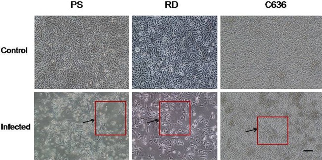
Susceptibility of different cell lines to Malsoor virus based on cytopathic effect (CPE) with 10× magnification. Cells were infected at a multiplicity of infection of 10. The areas of CPE were seen as microfoci of clearing in PS cells showing cellular changes (lower left panel) and distinct rounding of RD cells showing cellular changes on the 3rd p.i.d. (lower middle panel). There was no cytopathic effect in C6/36 cells on the 4th p.i.d. (lower right panel). The cultures were monitored for CPE until the 7th p.i.d. The arrows and the boxes outlined in red indicate areas of CPE. The magnification bar for all photomicrographs represents 1 μm.
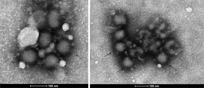
Representative transmission electron micrographs showing a typical negative stain showing bunyavirus particles in Vero CCL81 cells at the 4th p.i.d.. Scale bars are integrated into the micrograph. The arrows indicate representative bunyavirus particles showing distinct envelopes.
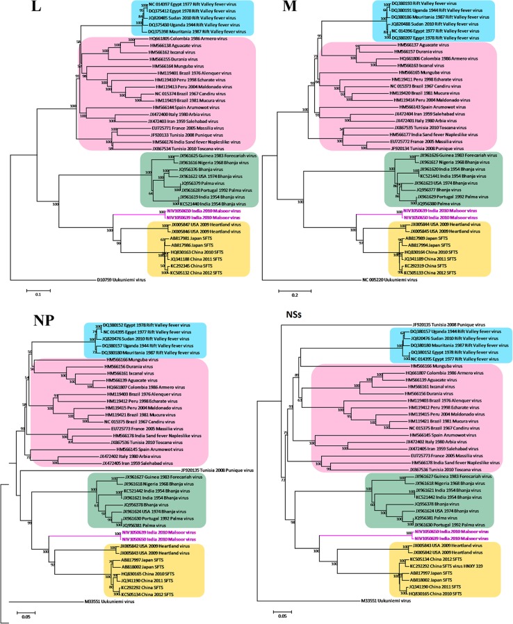
Phylogenetic analysis of N, NSs, M, and L segments of Malsoor virus. The phleboviruses and GenBank sequences used for phylogentic analysis of M, L, and S segments are as follows: Aguacate virus (HM566137, HM566138, HM566139), Alenquer virus (HM119402, HM119401, HM119403), Arbia virus (JX472401, JX472400, JX472402), Armero virus (HQ661806, HQ661805, HQ661807), Arumowot virus (HM566143, HM566144, HM566145), Bhanja virus (JQ956377, JQ956376, JQ956378), Bhanja virus (JX961617, JX961616, JX961618), Bhanja virus (JX961620, JX961619, JX961621), Bhanja virus (JX961623, JX961622, JX961624), Bhanja virus (KC521441, KC521440, KC521442), Candiru virus (NC_015373, NC_015374, NC_015375), Durania virus (HM566157, HM566155, HM566156), Echarate virus (HM119411, HM119410, HM119412), Forecariah virus (JX961626, JX961625, JX961627), Heartland virus (JX005844, JX005846, JX005842), Heartland virus (JX005845, JX005847, JX005843), Ixcanal virus (HM566163, HM566162, HM566161), Maldonado virus (HM119414, HM119413, HM119415), Massilia virus (EU725772, EU725771, EU725773), Mucura virus (HM119420, HM119419, HM119421), Munguba virus (HM566165, HM566164, HM566166), Palma virus (JQ956380, JQ956379, JQ956381), Palma virus (JX961629, JX961628, JX961630), phlebovirus JS24 (HQ830164, HQ830163, HQ830165), phlebovirus WCH/97/HN/China/2011 (JQ341189, JQ341188, JQ341190), Punique virus (JF920134, JF920133, JF920135), Rift Valley fever virus (RVFV) (DQ380186, DQ375398, DQ380180), RVFV (DQ380207, DQ375412, DQ380152), RVFV (DQ380193, DQ375430, DQ380157), RVFV (JQ820488, JQ820485, JQ820476), RVFV (NC_014396, NC_014397, NC_014395), Salehabad virus (JX472404, JX472403, JX472405), sand fever Naples-like virus (HM566177, HM566176, HM566178), severe fever with thrombocytopenia syndrome virus (SFTSV) (AB817989, AB817981, AB817997), SFTSV (AB817994, AB817986, AB818002), SFTSV (KC505133, KC505132, KC505134), SFTSV HNXY 319 (KC292319, KC292345, KC292292), Toscana virus (JX867535, JX867534, JX867536), and outgroup Uukuniemi virus (M33551, M17417, D10759).
References
-
- Pavri KM, Singh KR, Hollinger FB. 1971. Isolation of a new parainfluenza virus from a frugivorous bat, Rousettus leschenaulti, collected at Poona, India. Am. J. Trop. Med. Hyg. 20:125–130 - PubMed
-
- Newman SH, Field H, Epstein J, de Jong C. (ed). 2011. Investigating the role of bats in emerging zoonoses: balancing ecology, conservation and public health interest. FAO animal production and health manual no. 12. Food and Agriculture Organization of the United Nations, Rome, Italy: http://www.fao.org/docrep/014/i2407e/i2407e00.pdf
MeSH terms
Associated data
- Actions
- Actions
- Actions
- Actions
LinkOut - more resources
Full Text Sources
Other Literature Sources
Molecular Biology Databases
