. 2014 Mar;31(3):339-49.
doi: 10.1007/s10585-013-9632-3. Epub 2014 Jan 4.

CXCR4 antibody treatment suppresses metastatic spread to the lung of intratibial human osteosarcoma xenografts in mice

Affiliations

CXCR4 antibody treatment suppresses metastatic spread to the lung of intratibial human osteosarcoma xenografts in mice

Patrick Brennecke et al. Clin Exp Metastasis. 2014 Mar.

Abstract

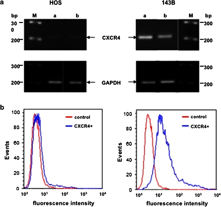
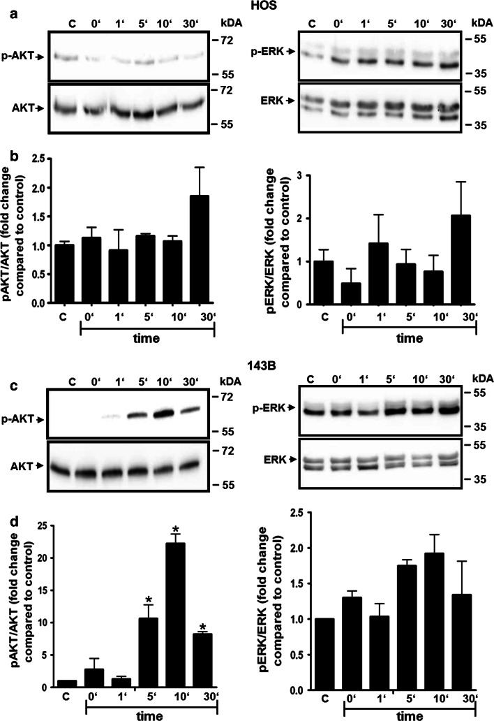
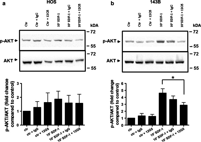
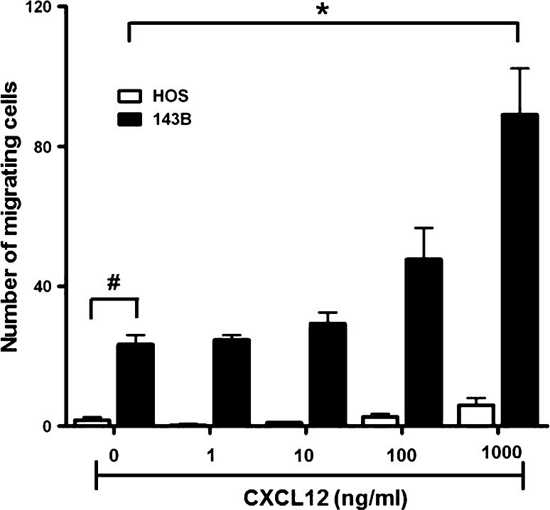
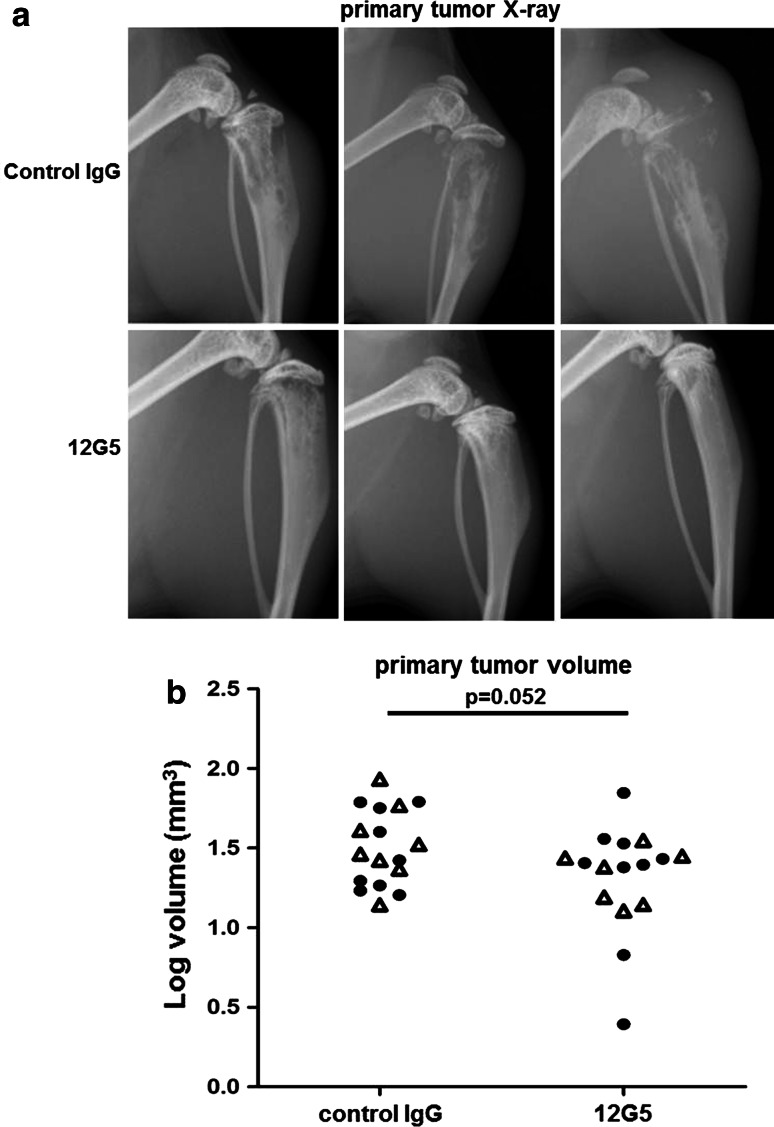
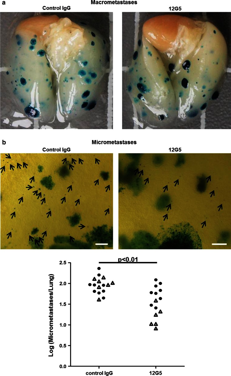
Current combined surgical and neo-adjuvant chemotherapy of primary metastatic osteosarcoma (OS) is ineffective, reflected by a 5-year survival rate of affected patients of less than 20 %. Studies in experimental OS metastasis models pointed to the CXCR4/CXCL12 homing axis as a novel target for OS metastasis-suppressive treatment. The present study investigated for the first time the CXCR4-blocking principle in a spontaneously metastasizing human 143B OS cell line-derived orthotopic xenograft mouse model. The highly metastatic 143B cells, unlike the parental non-metastatic HOS cells, express functional CXCR4 receptors at the cell surface, as revealed in this study by RT/PCR of gene transcripts, by FACS analysis with the monoclonal anti CXCR4 antibody 12G5 (mAb 12G5) and by CXCL12 time- and dose-dependent stimulation of AKT and ERK phosphorylation. A significantly (p < 0.05) higher CXCL12 dose-dependent chemotactic response of 143B compared to HOS cells in a Boyden chamber trans-well migration assay suggested a crucial role of the CXCL12/CXCR4 homing axis in 143B cell lung metastasis. Repetitive treatment of mice with 143B cell-derived intratibial tumors given intravenous bolus injections of mAb12G5 indeed inhibited significantly (p < 0.01) the number of X-gal-stainable lung micrometastases of lacZ-transduced 143B cells. Antibody treatment had also a mild inhibitory effect on primary tumor growth associated with remarkably less osteolysis, but it did not affect the number of developing lung macrometastases. In conclusion, these results demonstrate considerable potential of high-affinity CXCR4-blocking agents for OS tumor cell homing suppressive treatment in metastasizing OS complementary to current (neo)-adjuvant chemotherapy.


Figures

Fig. 1
Fig. 1
Expression of CXCR4 in the parental low metastatic human osteosarcoma HOS cell line and the highly metastatic 143B subline. a semi-quantitative RT/PCR analysis of Cxcr4 and Gapdh (reference) transcript levels in total RNA isolated from 50 % (a) and 100 % (b) confluent cells, M marker lanes. b FACS analysis of indicated cell lines processed for cell surface immunostaining of CXCR4 in the absence (control) or presence (CXCR4+) of primary 12G5 CXCR4 antibodies
Fig. 2
Fig. 2
Time-dependent stimulation of AKT and ERK phosphorylation by CXCL12 in HOS and 143B cells. a, b western blot analysis of extracts of HOS (a) and of 143B (c) cells incubated with 12.5 nM CXCL12 for indicated time periods. The levels of p-AKT and of total AKT (left panels) and of p-ERK and of total ERK (right panels) are shown. Data shown in a and c are representative for at least 3 independent experiments. b, d Quantitative analysis of the time-dependent stimulation of AKT (left) and ERK (right) phosphorylation in HOS (b) and143B (d) cells; Data indicate mean ± SEM (n = 3) levels of p-AKT and p-ERK normalized to total AKT and total ERK, respectively, at indicated time points; *p ≤ 0.05 compared to control (C)
Fig. 3
Fig. 3
AKT signaling inhibition by the blocking anti- CXCR4 antibody 12G5 in 143B and HOS cells. Shown are p-AKT and total AKT levels in HOS (a) and 143B (b) cells that were left untreated (ctr) or stimulated 10 min with SDF-1 (10′ SDF-1) and were pre-incubated with IgG (ctr + IgG and 10′ SDF-1 + IgG) or 12G5 (ctr + 12G5 and 10′ SDF-1 + 12G5) antibodies. Data indicate mean ± SEM (n = 3) levels of p-AKT normalized to total AKT (*p ≤ 0.05 compared to control 10′ SDF-1 stimulation)
Fig. 4
Fig. 4
CXCL12 dose-dependent CXCR4-mediated chemotaxis of HOS and 143B cells. Cells were seeded in 48-well Boyden chambers on membranes of 8 μm pore size and chemotaxis to indicated concentrations of CXCL12 added to the medium in the bottom chamber was allowed for 4 h. Non-migrating cells adhering to the upper surface of the membrane were removed and cells that had migrated across the membrane and adhered to the bottom surface were fixed, stained with Diff Quick® and counted under the microscope. *p  0.05 compared to 143B cells incubated in the absence of CXCL12; # p  0.05 compared to HOS cells in the absence of CXCL12
Fig. 5
Fig. 5
Effects of repetitive systemic administration of 12G5 CXCR4-blocking antibodies on intratibial primary tumor development of 143B-lacZ osteosarcoma cell xenografts. a Representative X-ray images of 143B-lacZ cell-derived tumors in severe combined immunodeficient mice treated with control mouse IgG (top panels) or with 12G5 CXCR4 antibodies (bottom panels). b Primary tumor volumes calculated from caliper measurements of tumor bearing tibiae in control mouse IgG- or 12G5 antibody-treated mice. The results are from two independent experiments with 7 (first experiment) and 6 (second treatment) (open triangle) and 9 (filled circle) mice per indicated group
Fig. 6
Fig. 6
Micrometastasis-inhibiting effects of repetitive administration of CXCR4-blocking 12G5 antibodies in mice with intratibial 143B-lacZ cell-derived tumors. a Representative images of whole mounts of lungs of mice treated with mouse IgG (left) or with 12G5 CXCR4 antibodies (right). X-gal stained metastases appear in blue. b Microscopic images of representative areas of lung whole mounts (4-fold magnification, size bar = 10 μm) of 143B-lacZ cell tumor-bearing mice treated with mouse IgG (left) or with 12G5 CXCR4 antibodies (right). Arrows point to X-gal-stained micrometastases in blue. The total numbers of micrometastases per lung of indicated mice treated with mouse IgG or with 12G5 antibodies are shown in the bottom panel. The results were obtained in two independent experiments with 7 (open triangle) and 9 (filled circle) mice per indicated group. p < 0.01 was considered as statistically significant

References

    1. Federman N, Bernthal N, Eilber FC, Tap WD. The multidisciplinary management of osteosarcoma. Curr Treat Options Oncol. 2009;10(1–2):82–93. doi: 10.1007/s11864-009-0087-3. - DOI - PubMed
    1. Picci P. Osteosarcoma (osteogenic sarcoma) Orphanet J Rare Dis. 2007;2:6. doi: 10.1186/1750-1172-2-6. - DOI - PMC - PubMed
    1. Carrle D, Bielack SS. Current strategies of chemotherapy in osteosarcoma. Int Orthop. 2006;30(6):445–451. doi: 10.1007/s00264-006-0192-x. - DOI - PMC - PubMed
    1. Mercuri M, Capanna R, Manfrini M, Bacci G, Picci P, Ruggieri P, Ferruzzi A, Ferraro A, Donati D, Biagini R, et al. The management of malignant bone tumors in children and adolescents. Clin Orthop Relat Res. 1991;264:156–168. - PubMed
    1. Jaffe N, Carrasco H, Raymond K, Ayala A, Eftekhari F. Can cure in patients with osteosarcoma be achieved exclusively with chemotherapy and abrogation of surgery? Cancer. 2002;95(10):2202–2210. doi: 10.1002/cncr.10944. - DOI - PubMed

Publication types

MeSH terms