. 2014 Jun;111(6):1222-32.
doi: 10.1002/bit.25180. Epub 2014 Jan 22.

Synthetic small intestinal scaffolds for improved studies of intestinal differentiation

Affiliations

Synthetic small intestinal scaffolds for improved studies of intestinal differentiation

Cait M Costello et al. Biotechnol Bioeng. 2014 Jun.

Abstract

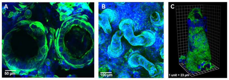
In vitro intestinal models can provide new insights into small intestinal function, including cellular growth and proliferation mechanisms, drug absorption capabilities, and host-microbial interactions. These models are typically formed with cells cultured on 2D scaffolds or transwell inserts, but it is widely understood that epithelial cells cultured in 3D environments exhibit different phenotypes that are more reflective of native tissue. Our focus was to develop a porous, synthetic 3D tissue scaffold with villous features that could support the culture of epithelial cell types to mimic the natural microenvironment of the small intestine. We demonstrated that our scaffold could support the co-culture of Caco-2 cells with a mucus-producing cell line, HT29-MTX, as well as small intestinal crypts from mice for extended periods. By recreating the surface topography with accurately sized intestinal villi, we enable cellular differentiation along the villous axis in a similar manner to native intestines. In addition, we show that the biochemical microenvironments of the intestine can be further simulated via a combination of apical and basolateral feeding of intestinal cell types cultured on the 3D models.

Keywords: caco-2; cadherin; egf; ht29; lysozyme; muc-2.


Figures

Figure 1
Figure 1
Schematic representation of porous PLGA intestinal scaffold formation. Laser ablation is used to create an array of 500 μm deep holes in a PMMA (A). PDMS replicas fabricated to produce a PDMS intestinal scaffold template (B and C). Agarose replicas are made to enable improved detachment of the final PLGA scaffolds (D and E). High PLGA/low porogen solution under vacuum to forms initial villous layer (F) with additional polymer layer (low PLGA/high porogen) added to increase the porosity. Total thickness of the base measures approximately 500 μm (G). Scaffolds are then frozen at −20°C, immersed in pre-cooled ethanol to extract the solvent, then incubated in warm water to dissolve the porogen.
Figure 2
Figure 2
SEM images of PLGA scaffolds showing non-porous PLGA scaffolds (A) and PLGA scaffolds made porous by porogen leaching and solvent extraction (B) Cell-free resistance of PLGA scaffolds made porous through a combination of solvent extraction and porogen leaching, or porogen leaching in isolation (C).
Figure 3
Figure 3
Confocal microscopy of a PLGA scaffold co-cultured with Caco-2 and HT29-MTX with staining for nuclei (blue) and actin (green). Twenty times magnification shows partial coverage of villi by cells after 4 days (A), with full coverage of cells over a section of scaffold from the base to the tips after 7 days (B) and 3D rendering shows full coverage of cells on an individual villus measuring 500 μm (C).
Figure 4
Figure 4
Confocal imaging of whole 3D scaffolds with staining for nuclei (blue), actin (green). Z stack Images show change in cell morphology after 7 days (A), 14 days (B), and 21 days co-culture (C). Cryo-sectioned scaffold slices, with staining for alkaline phosphatase (red), show an increase in enzyme expression between 7, 14 and 21 days (D–F, respectively).
Figure 5
Figure 5
TEER values (A) and alkaline phosphatase activity (B) of co-cultures on PLGA scaffolds and transwell inserts with and without EGF stimulation basolaterally. Statistics were performed using a paired t-test (P <0.01).
Figure 6
Figure 6
Confocal imaging of scaffold slice (A) and 3D z stacks of scaffolds with staining for nuclei (blue), and mucus (red). Images were taken after 21 days. Three dimensional scanning shows the tip (B), middle (C) and bottom of an individual villus (D). Mucus production over 21 days was also assessed using an ELLA with wheat germ agglutinin to bind and quantify mucus. Results are expressed as ng/mL mucus in reference to a standard curve, and normalized to cell density (mucus production per 100,000 cells (P <0.01).
Figure 7
Figure 7
Confocal microscopy of a PLGA scaffold co-cultured with mouse small intestinal crypts for 5 days with staining for nuclei (blue), e-cadherin (green) lysozyme and MUC-2 (red). 10× magnification shows full coverage of villi by crypt cells that have differentiated into enterocytes, paneth and goblet cells. Arrows point to paneth cells stained for lysozyme that remain at the base (A) and goblet cells stained for MUC-2, which have migrated up the villi (B).

References

    1. Andersson AS, Brink J, Lidberg U, Sutherland DS. Influence of systematically varied nanoscale topography on the morphology of epithelial cells. IEEE Trans Nanobiosci. 2003;2(2):49–57. - PubMed
    1. Anselme K, Bigerelle M. Role of materials surface topography on mammalian cell response. Int Mater Rev. 2011;56(4):243–266.
    1. Atreya R, Mudter J, Finotto S, Mullberg J, Jostock T, Wirtz S, Schutz M, Bartsch B, Holtmann M, Becker C, Strand D, Czaja J, Schlaak JF, Lehr HA, Autschbach F, Schurmann G, Nishimoto N, Yoshizaki K, Ito H, Kishimoto T, Galle PR, Rose-John S, Neurath MF. Blockade of interleukin 6 trans signaling suppresses T-cell resistance against apoptosis in chronic intestinal inflammation: Evidence in Crohn disease and experimental colitis in vivo. Nat Med. 2000;6(5):583–588. - PubMed
    1. Ayabe T, Satchell DP, Pesendorfer P, Tanabe H, Wilson CL, Hagen SJ, Ouellette AJ. Activation of Paneth cell alpha-defensins in mouse small intestine. J Biol Chem. 2002;277(7):5219–5228. - PubMed
    1. Ayabe T, Satchell DP, Wilson CL, Parks WC, Selsted ME, Ouellette AJ. Secretion of microbicidal alpha-defensins by intestinal Paneth cells in response to bacteria. Nat Immunol. 2000;1(2):113–118. - PubMed

Publication types