. 2014 Jun;59(6):2263-75.
doi: 10.1002/hep.26993. Epub 2014 Apr 25.

Cholangiocyte senescence by way of N-ras activation is a characteristic of primary sclerosing cholangitis

Affiliations

Cholangiocyte senescence by way of N-ras activation is a characteristic of primary sclerosing cholangitis

James H Tabibian et al. Hepatology. 2014 Jun.

Abstract

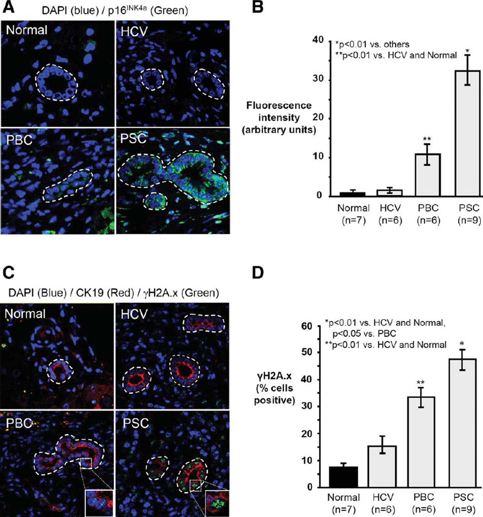
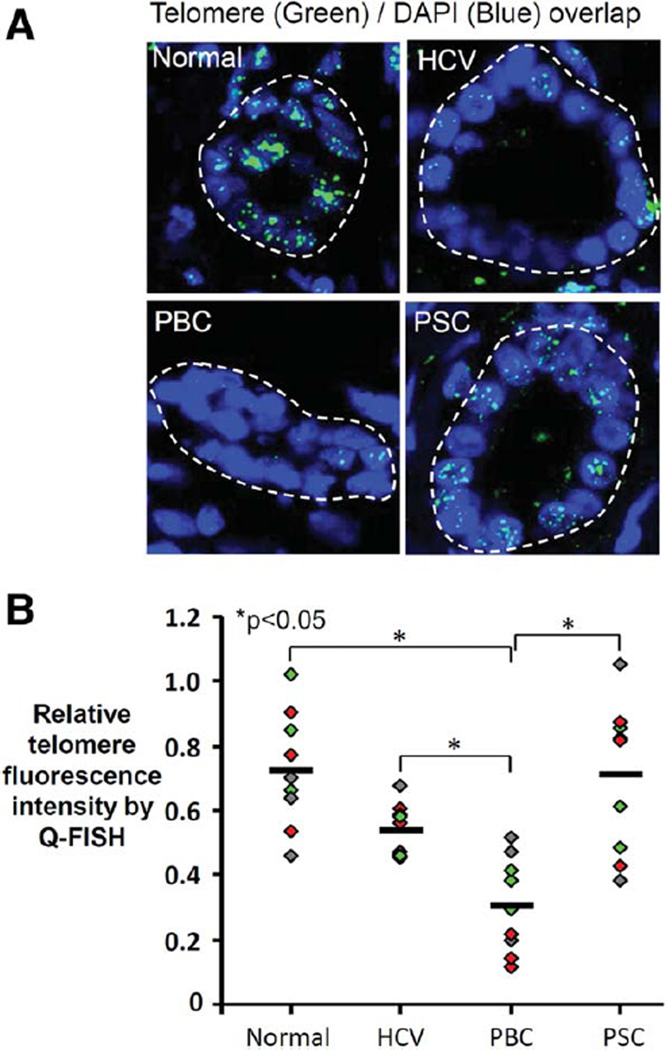
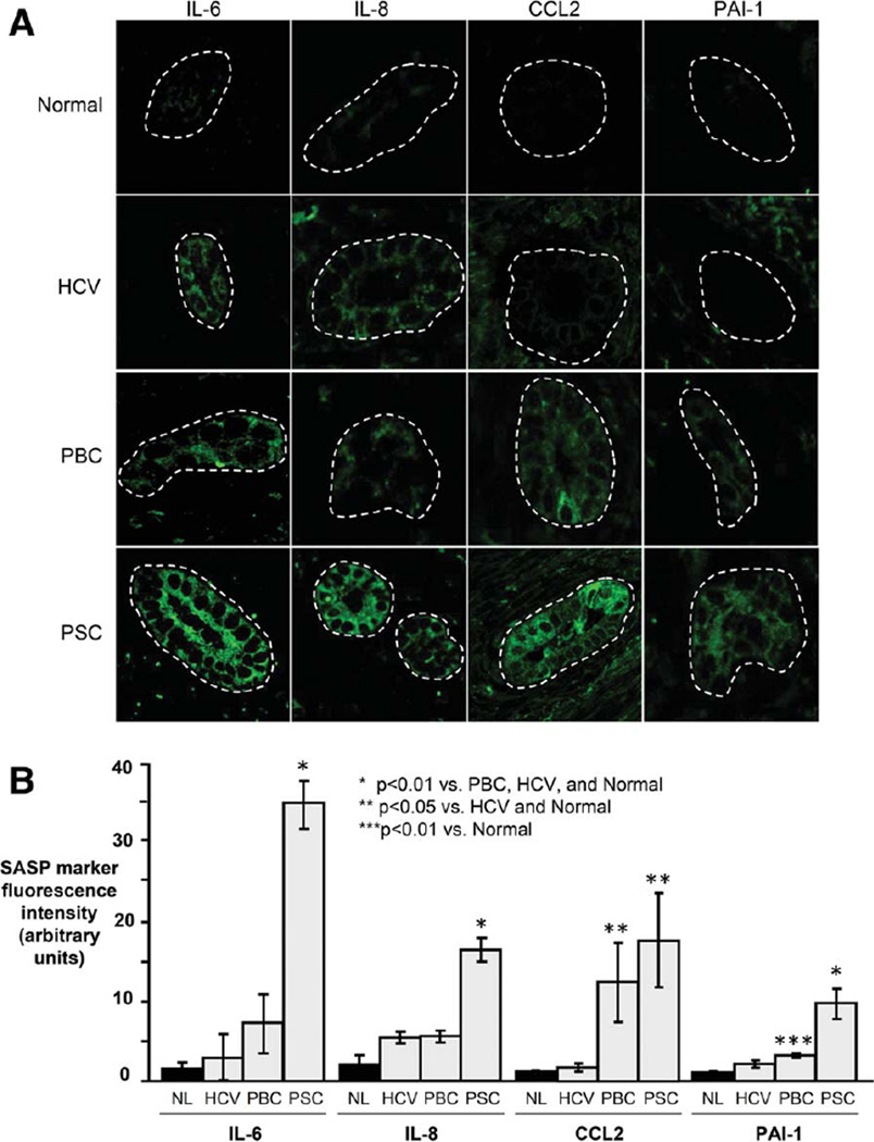
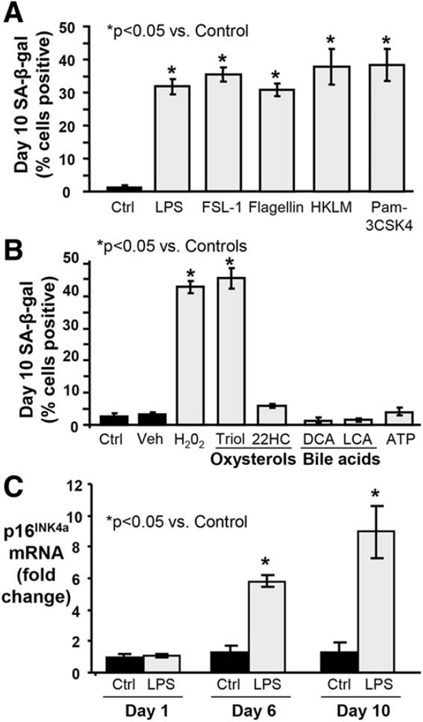
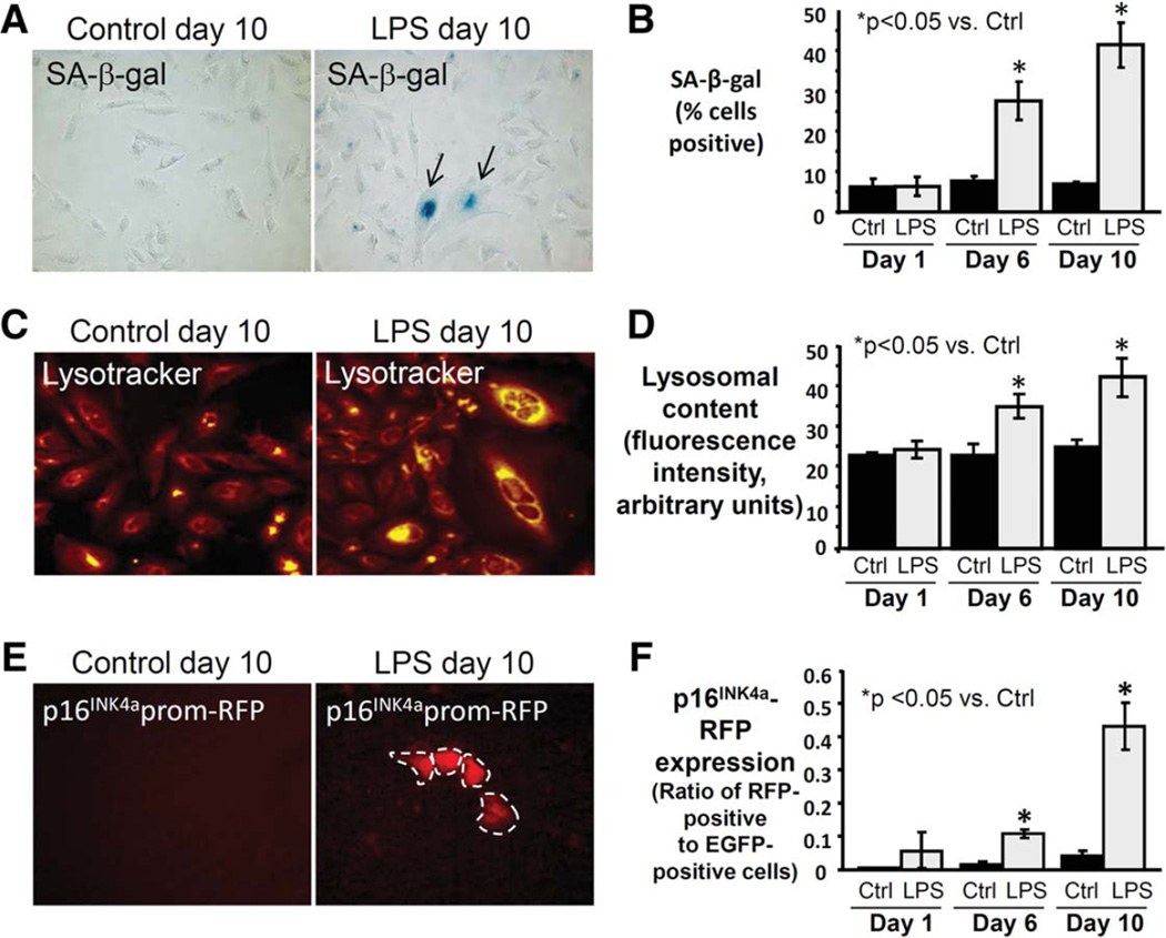
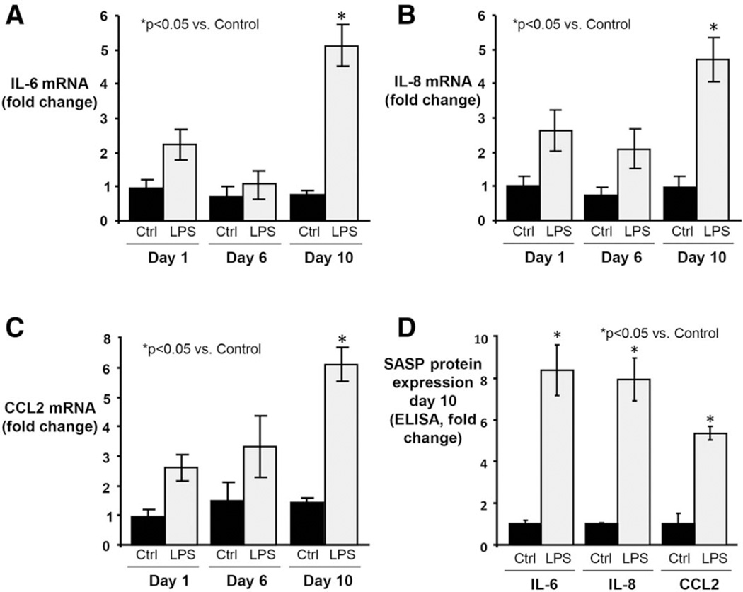
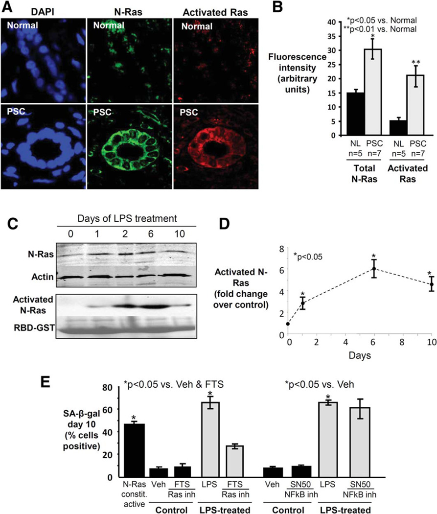
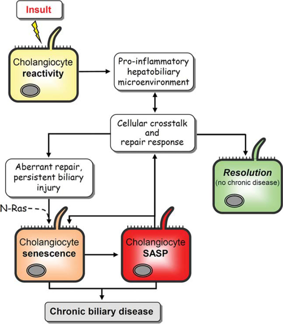
Primary sclerosing cholangitis (PSC) is an incurable cholangiopathy of unknown etiopathogenesis. Here we tested the hypothesis that cholangiocyte senescence is a pathophysiologically important phenotype in PSC. We assessed markers of cellular senescence and senescence-associated secretory phenotype (SASP) in livers of patients with PSC, primary biliary cirrhosis, hepatitis C, and in normals by fluorescent in situ hybridization (FISH) and immunofluorescence microscopy (IFM). We tested whether endogenous and exogenous biliary constituents affect senescence and SASP in cultured human cholangiocytes. We determined in coculture whether senescent cholangiocytes induce senescence in bystander cholangiocytes. Finally, we explored signaling mechanisms involved in cholangiocyte senescence and SASP. In vivo, PSC cholangiocytes expressed significantly more senescence-associated p16(INK4a) and γH2A.x compared to the other three conditions; expression of profibroinflammatory SASP components (i.e., IL-6, IL-8, CCL2, PAI-1) was also highest in PSC cholangiocytes. In vitro, several biologically relevant endogenous (e.g., cholestane 3,5,6 oxysterol) and exogenous (e.g., lipopolysaccharide) molecules normally present in bile induced cholangiocyte senescence and SASP. Furthermore, experimentally induced senescent human cholangiocytes caused senescence in bystander cholangiocytes. N-Ras, a known inducer of senescence, was increased in PSC cholangiocytes and in experimentally induced senescent cultured cholangiocytes; inhibition of Ras abrogated experimentally induced senescence and SASP.

Conclusion: Cholangiocyte senescence induced by biliary constituents by way of N-Ras activation is an important pathogenic mechanism in PSC. Pharmacologic inhibition of N-Ras with a resultant reduction in cholangiocyte senescence and SASP is a new therapeutic approach for PSC.


Conflict of interest statement

Potential conflict of interest: Nothing to report.

Figures

Fig. 1
Fig. 1
Cholangiocytes in PSC liver exhibit increased markers of cellular senescence. (A) Representative images of p16INK4A mRNA FISH (p16Ink4A probe = green; DAPI = blue). (B) Semiquantitative analysis of fluorescence intensity demonstrates significantly increased cholangiocyte p16INK4A in PSC compared to other conditions. (C) Representative images of γH2A.x immunofluorescence (γH2A.x = green; DAPI = blue). (D) Quantitation of γH2A.x foci demonstrates a higher proportion of γH2A.x-positive cholangiocytes in PSC compared to other conditions.
Fig. 2
Fig. 2
Telomere length is preserved in PSC cholangiocytes. (A) Representative confocal fluorescence images of Tel-C-FITC FISH performed on liver sections to assess cholangiocyte telomere length. (B) Cholangiocyte telomere fluorescence intensity is similar in PSC and normal liver but significantly decreased in PBC. Diamonds represent the average relative telomere fluorescence intensity of bile ducts in a randomly selected liver section (three sections assessed in each of three patients, each patient shown in a different color); horizontal line represents the average of the three patients in each condition. For analysis, three average values were used for each patient based on the three randomly selected liver sections that were assessed. If per-patient average values are used instead of per-section average values, the difference between HCV and PBC loses statistical significance, while the difference between PSC and PBC and the difference between normal and PBC retain statistical significance (both P < 0.05).
Fig. 3
Fig. 3
Cholangiocytes in PSC liver exhibit increased expression of SASP components. (A) Representative images of IL-6, IL-8, CCL2, and PAI-1 immunofluorescence in liver sections. (B) Semi-quantitative analysis of fluorescence intensity (presented as arbitrary units) demonstrates significantly increased cholangiocyte expression of SASP components in PSC.
Fig. 4
Fig. 4
In vitro model of stress-induced NHC senescence by persistent treatment with exogenous and endogenous insults. NHCs were exposed to exogenous (i.e., microbially derived) and endogenous stressors over the course of 10 days. (A) The proportion of SA-β-gal-positive NHCs increases following 10-day treatment with exogenous stressors, indicating induction of senescence. FSL-1, synthetic diacylated lipoprotein; HKLM, heat-killed L. monocytogenes; Pam3CSK4, synthetic triacylated lipoprotein. (B) The proportion of SA-β-gal-positive NHCs increases following 10-day treatment with (some) endogenous stressors, indicating induction of senescence. Veh, vehicle (ethanol) for Triol (cholestane-3β, 5α, 6α-triol); 22HC, 22-hydroxycholesterol; DCA, deoxycholic acid; LCA, lithocholic acid; ATP, adenosine triphosphate. (C) Persistent LPS treatment induces significantly increased p16INK4a mRNA expression at day 6 day and a further increase at day 10 compared to control cells.
Fig. 5
Fig. 5
Persistent treatment with LPS induces expression of multiple senescence markers in NHCs. (A) Representative brightfield microscopy images at day 10 demonstrating increased LPS-induced SA-β-gal and cytologic features of senescence (larger size, squamoid appearance). (B) SA-β-gal quantitation reveals significantly increased proportion of SA-β-gal-positive NHCs following 6 and 10 days of LPS treatment. (C) Representative images at day 10 demonstrating increased LPS-induced lysosomal content. (D) Quantitation of lysosomal content reveals a significant increase in lysosomal content at day 6 and a further increase at day 10 compared to control. (E) Representative images at day 10 demonstrating LPS-induced expression of p16INK4a promoter-driven reporter (RFP). (F) Quantitation of p16INK4a (presented as the ratio of RFP to EGFP-positive cells) confirms a significant increase at day 6 and a further increase at day 10 compared to control.
Fig. 6
Fig. 6
Persistent LPS treatment of NHCs induces expression of SASP components. (A) IL-6 mRNA expression was assessed by qPCR at 1, 6, and 10 days of LPS treatment. By day 10, expression is significantly elevated (~5-fold > control; ~2-fold > 1 day LPS-treatment). (B) IL-8 mRNA expression is significantly increased by day 10 (~5-fold > control; ~1.5-fold > 1 day LPS-treatment). The delayed response observed in IL-8 (as well as IL-6) expression following an initial acute response is consistent with the time-dependent induction of senescence as demonstrated in other cell types and culture models. (C) CCL2 mRNA expression is significantly increased by day 10 (~4.7-fold > control; ~1.9-fold > 1 day LPS-treatment). (D) ELISA of conditioned media from 10-day LPS-treated NHCs demonstrating significantly increased IL-6, IL-8, and CCL2 protein expression compared to control.
Fig. 7
Fig. 7
Ras is activated in cholangiocytes in PSC liver, and Ras activation is involved in LPS-induced NHC senescence. (A) Representative con-focal immunofluorescence images for N-Ras (green) and activated Ras (red) (DAPI = blue). Cholangiocyte N-Ras colocalizes with activated Ras in PSC liver, suggesting that N-Ras is not only expressed, but also activated in PSC cholangiocytes. (B) Semiquantitative analysis of fluorescence demonstrates significantly increased N-Ras and activated Ras in PSC cholangiocytes compared to normal. (C) LPS treatment of NHCs induces persistent N-Ras activation. Western blot demonstrates cholangiocyte N-Ras expression, and an RBD-GST pulldown demonstrates LPS-induced Ras activation over the course of 10 days (Ponceau Red used to stain total RBD-GST, loading control). (D) Densitometry on the activated N-Ras blot at each time point. (E) Ras, but not NF-κB inhibition, decreases LPS-induced cholangiocyte senescence. NHCs were cultured in the presence or absence of LPS and a Ras (FTS) or NF-κB (SN50) inhibitor. Cholangiocyte senescence, determined by proportion of SA-β-gal-positive cholangiocytes, is abrogated by Ras inhibition.
Fig. 8
Fig. 8
Conceptual framework of PSC pathogenesis. In this working model, the cholangiocyte is exposed to endogenous and/or exogenous insults to which it responds through up-regulation of proinflammatory mediators as characteristic of a “reactive cholangiocyte.” This process initiates intricate crosstalk between a variety of resident and recruited cells, including hepatocytes, progenitor cells, fibroblasts, and leukocytes, in an attempt to resolve injury and repair the biliary epithelium. In immunogenetically susceptible individuals, the proinflammatory response of and injury to cholangiocytes do not resolve. Instead, as a result of aberrant genomic and cellular repair, we speculate, based on our data and current hypotheses, that there is induction of N-Ras mediated, insult-induced cholangiocyte senescence. Senescent cholangiocytes can then progress to SASP, a potentially pathologic state characterized by hypersecretion of proinflammatory cytokines (e.g., IL-6) and chemokines (e.g., IL-8) and profibrotic mediators (e.g., PAI-1), as shown in the present study. By way of these secreted mediators, SASP cells have been shown to alter their microenvironment, reinforce the senescent phenotype, and exacerbate injurious fibroinflammatory responses which, in the liver, results in progressive injury and ultimately chronic biliary disease (i.e., PSC).

References

    1. Tabibian JH, Lindor KD. Primary sclerosing cholangitis: a review and update on therapeutic developments. Expert Rev Gastroenterol Hepatol. 2013;7:103–114. - PubMed
    1. O’Hara SP, Tabibian JH, Splinter PL, Larusso NF. The dynamic biliary epithelia: molecules, pathways, and disease. J Hepatol. 2013;58:575–582. - PMC - PubMed
    1. Wiesner RH, Grambsch PM, Dickson ER, Ludwig J, MacCarty RL, Hunter EB, et al. Primary sclerosing cholangitis: natural history, prognostic factors and survival analysis. Hepatology. 1989;10:430–436. - PubMed
    1. Tabibian JH, Lindor KD. Challenges of cholangiocarcinoma detection in patients with primary sclerosing cholangitis. J Anal Oncol. 2012;1:50–55. - PMC - PubMed
    1. Alabraba E, Nightingale P, Gunson B, Hubscher S, Olliff S, Mirza D, et al. A re-evaluation of the risk factors for the recurrence of primary sclerosing cholangitis in liver allografts. Liver Transpl. 2009;15:330–340. - PubMed

Publication types