. 2013 Dec;11(12):e1001750.
doi: 10.1371/journal.pbio.1001750. Epub 2013 Dec 31.

The velvet family of fungal regulators contains a DNA-binding domain structurally similar to NF-κB

Affiliations

The velvet family of fungal regulators contains a DNA-binding domain structurally similar to NF-κB

Yasar Luqman Ahmed et al. PLoS Biol. 2013 Dec.

Erratum in

  • PLoS Biol. 2014 Apr;12(4):e1001849

Abstract

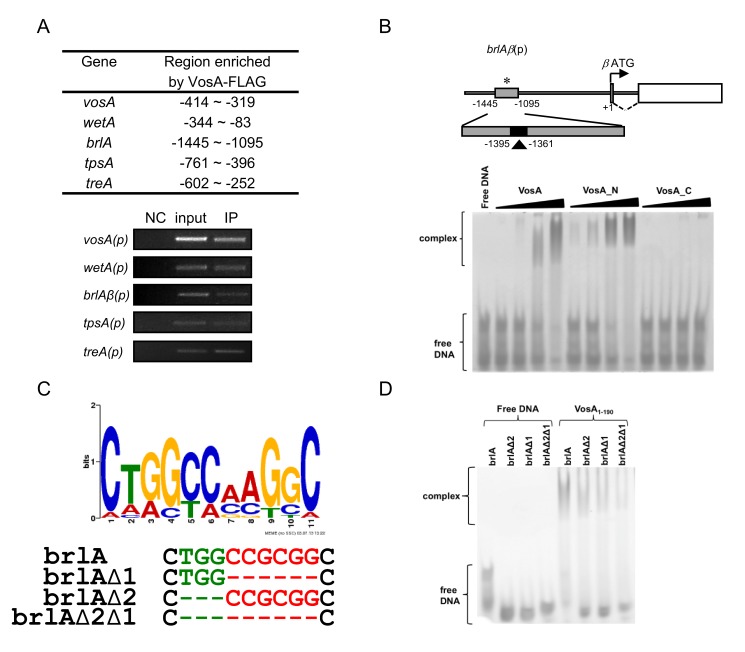
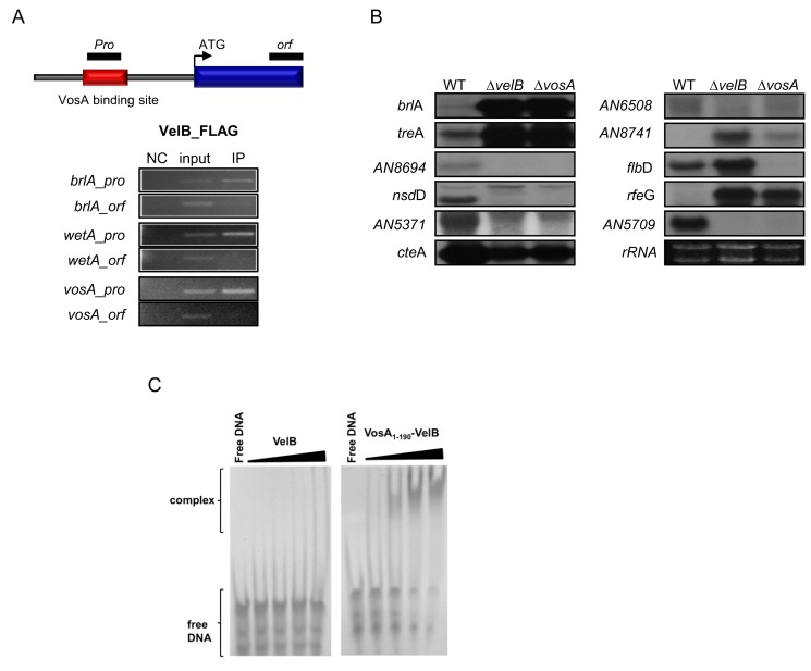
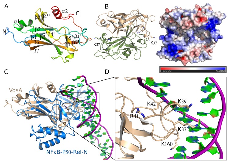
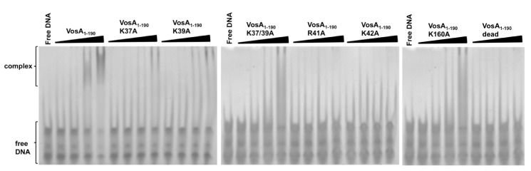
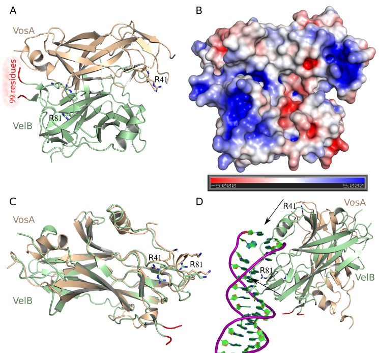
Morphological development of fungi and their combined production of secondary metabolites are both acting in defence and protection. These processes are mainly coordinated by velvet regulators, which contain a yet functionally and structurally uncharacterized velvet domain. Here we demonstrate that the velvet domain of VosA is a novel DNA-binding motif that specifically recognizes an 11-nucleotide consensus sequence consisting of two motifs in the promoters of key developmental regulatory genes. The crystal structure analysis of the VosA velvet domain revealed an unforeseen structural similarity with the Rel homology domain (RHD) of the mammalian transcription factor NF-κB. Based on this structural similarity several conserved amino acid residues present in all velvet domains have been identified and shown to be essential for the DNA binding ability of VosA. The velvet domain is also involved in dimer formation as seen in the solved crystal structures of the VosA homodimer and the VosA-VelB heterodimer. These findings suggest that defence mechanisms of both fungi and animals might be governed by structurally related DNA-binding transcription factors.


Conflict of interest statement

The authors have declared that no competing interests exist.

Figures

Figure 1
Figure 1. VosA binds DNA specifically.
(A) Selected regions enriched by VosA-FLAG ChIP on chip. The promoter regions of brlA, wetA, vosA, tpsA, and treA enriched by VosA-FLAG ChIP are shown (the start codon ATG is +1). Results of VosA-ChIP-PCR, the PCR amplicons separated on a 2% agarose gel, are shown in the bottom panel. The input DNA before immuno-precipitation (IP) was used as a positive control (input). The chromatin extract being incubated with bead only (without anti-FLAG antibody) was used as a negative control (NC). (B) Schematic presentation of the promoter region of brlA. The gray box (−1,095∼1,445 region of brlA(p), marked by *) represents the ChIP-PCR amplified region shown in (A), and the filled box (marked by arrowhead) represents a 35 bp fragment used in EMSA. EMSA using serially diluted VosA proteins and the 35 bp DNA probe of the brlA promoter (OHS301/302). DNA and protein were used in the molar ratios 1∶0.3, 1∶1, 1∶3, and 1∶9. VosA, full-length VosA; VosA_N, truncated VosA (residues 1–216); VosA_C, truncated VosA (residues 217–430). (C) The consensus DNA sequence predicted to be recognized by VosA is shown. Two cores of this motif were deleted as shown (green, red) and these probes were used for additional EMSA. (D) EMSA using the crystallized VosA1–190 (VosA_cryst) with wild-type and mutated DNA probes of the brlA promoter. In the mutated versions of the DNA, the predicted VosA binding motifs 1 and 2 were deleted (brlA, wild-type DNA (OHS301/302); brlAΔ2, DNA with deleted core 2 (JG636/637); brlAΔ1, DNA with deleted core 1 (JG638/639); brlAΔ2Δ1, DNA with deleted cores 2 and 1 (JG640/641)).
Figure 2
Figure 2. Regulatory roles of VelB and VosA1–190-VelB complex.
(A) Schematic presentation of the promoter region of a typical vosA target gene. The red box represents the region, which was enriched by VosA-ChIP (upper). Black boxes represent two ChIP-PCR target regions shown at the bottom. Results of VelB-ChIP-PCR, the PCR amplicons separated on a 2% agarose gel, are shown (bottom). The input DNA before immunoprecipitation (IP) was used as a positive control (input). The chromatin extract being incubated with bead only (without anti-FLAG antibody) was used as a negative control (NC). (B) mRNA levels of the genes that are predicted to be under the direct regulatory control of VosA in the conidia of wt (wild-type; FGSC4), ΔvelB (THS16.1), and ΔvosA (THS15.1) strains. Equal loading of total RNA was confirmed by ethidium bromide staining of rRNA. (C) EMSA using the VosA1–190-VelB heterodimer and VelB with the 35 bp DNA probe of the brlA promoter (OHS301/302). DNA and protein were used in the molar ratios 1∶0.3, 1∶1, 1∶3, and 1∶9.
Figure 3
Figure 3. Crystal structure of VosA1–190.
(A) Structure of the velvet domain of VosA. Amino acid residues 1–185 of VosA1–190 from A. nidulans fold into a seven-stranded β-sandwich and two α-helices. (B) Structure of the VosA homodimer. The two monomers are related by a crystallographic 2-fold symmetry axis. Loop A is indicated by the labelling of K37. The electrostatic surface potential of the VosA1–190 homodimer indicates the binding surface for DNA. Surface representation coloured according to the electrostatic potential ranging from red (−5 kBT/e) through white (0 kBT/e) to blue (+5 kBT/e). (C) Superposition of the VosA velvet domain and the Rel-N domain of NF-κB bound to DNA . (D) Lys and Arg residues of VosA1–190 that are predicted to be involved in DNA binding as deduced by the superposition VosA1–190 onto the NF-κB-DNA complex structure (loop B is indicated by the labelling of K160).
Figure 4
Figure 4. EMSA of the wild-type and mutated VosA1–190 proteins.
EMSA data using wild-type and mutated VosA1–190 with the 35 bp DNA probe of the brlA promoter (OHS301/302) containing the predicted VosA-binding sequence are shown. The “dead” mutant contains four substitutions (K37A, K39A, R41A, and K42A). DNA and protein were used in the molar ratios 1∶0.3, 1∶1, 1∶3, and 1∶9. Free DNA without protein was used as negative control.
Figure 5
Figure 5. Crystal structure of the VosA1–190-VelB complex.
(A) Structure of the VosA1–190-VelB heterodimer formed by the velvet domains of both proteins. Residues 1–7, 37–39, and 161–190 of VosA are not defined in the electron density map, as well as 99 residues inserted into the velvet domain of VelB. These 99 residues are predicted to be an intrinsically disordered region that is inserted into VelB in the gap highlighted by the two orange coloured disconnected ends of the VelB polypeptide chain. (B) The electrostatic surface potential of the VosA1–190-VelB heterodimer indicates a binding surface for DNA similar to that of the VosA homodimer. Surface representation coloured according to the electrostatic potential ranging from red (−5 kBT/e) through white (0 kBT/e) to blue (+5 kBT/e). (C) Superposition of the velvet domains of VosA (in blue) and VelB (in green). The position of the missing 99-residue insertion of the VelB velvet domain is highlighted in orange. (D) Proposed model for DNA-binding of the VosA1–190-VelB heterodimer based on the NF-κB-DNA complex structure . The arrows show the location of basic residues involved in DNA-binding. Contacts have been optimized for the VelB-subunit leading to a nonoptimal position of the VosA binding site. Hence, some bending of the dsDNA might be introduced upon binding of the heterodimer.

Comment in

  • The velvet underground emerges.
    Roberts RG. Roberts RG. PLoS Biol. 2013 Dec;11(12):e1001751. doi: 10.1371/journal.pbio.1001751. Epub 2013 Dec 31. PLoS Biol. 2013. PMID: 24391471 Free PMC article. No abstract available.

References

    1. Baldauf SL, Palmer JD (1993) Animals and fungi are each other's closest relatives: congruent evidence from multiple proteins. Proc Natl Acad Sci USA 90: 11558–11562. - PMC - PubMed
    1. Wainright PO, Hinkle G, Sogin ML, Stickel SK (1993) Monophyletic origins of the metazoa: an evolutionary link with fungi. Science 260: 340–342. - PubMed
    1. O'Dea E, Hoffmann A (2010) The regulatory logic of the NF-kappaB signaling system. Cold Spring Harb Perspect Biol 2: a000216. - PMC - PubMed
    1. Oeckinghaus A, Hayden MS, Ghosh S (2011) Crosstalk in NF-kappaB signaling pathways. Nat Immunol 12: 695–708. - PubMed
    1. Calvo AM, Wilson RA, Bok JW, Keller NP (2002) Relationship between secondary metabolism and fungal development. Microbiol Mol Biol Rev 66: 447–459. - PMC - PubMed

Publication types

MeSH terms

LinkOut - more resources