. 2014 Jan;10(1):e1003844.
doi: 10.1371/journal.ppat.1003844. Epub 2014 Jan 2.

Inflammation fuels colicin Ib-dependent competition of Salmonella serovar Typhimurium and E. coli in enterobacterial blooms

Affiliations

Inflammation fuels colicin Ib-dependent competition of Salmonella serovar Typhimurium and E. coli in enterobacterial blooms

Lubov Petkova Nedialkova et al. PLoS Pathog. 2014 Jan.

Abstract

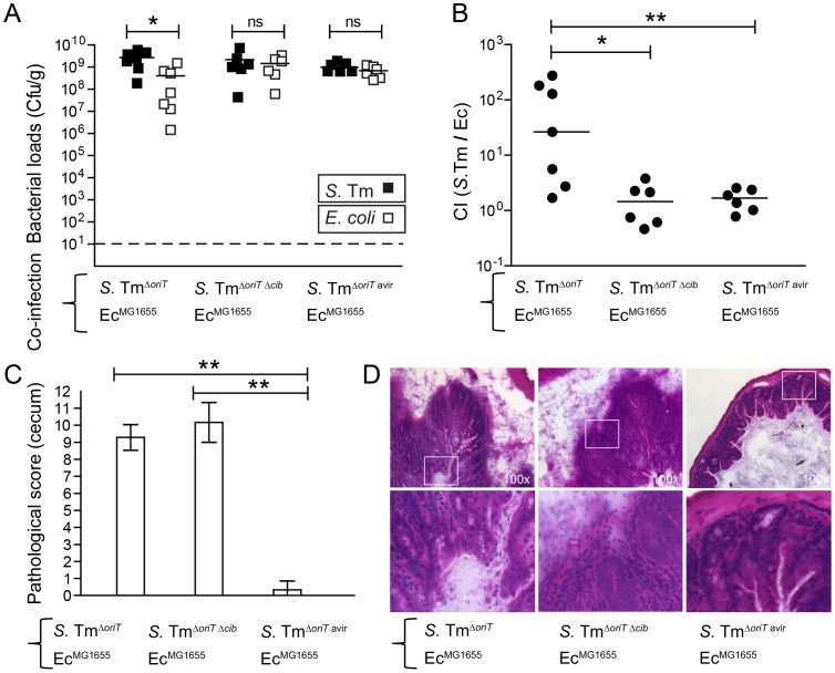
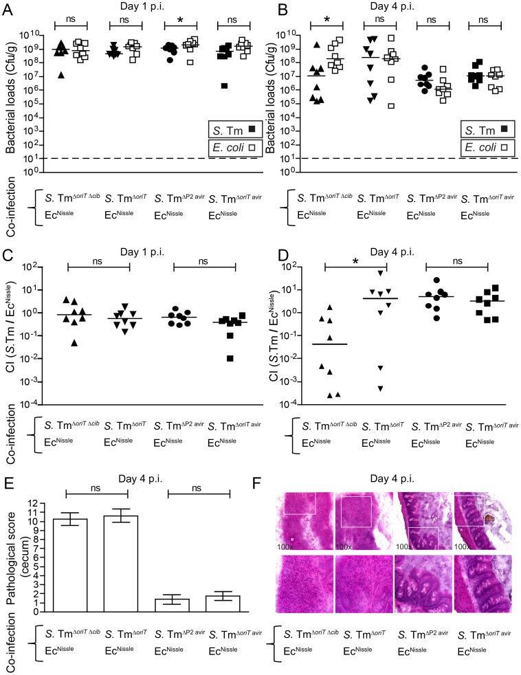
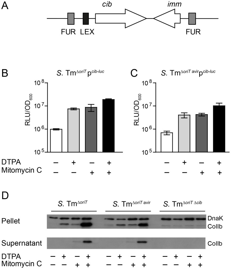
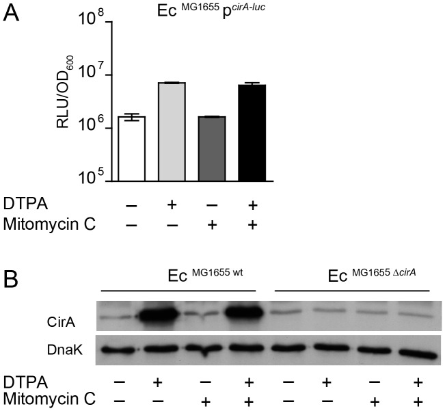
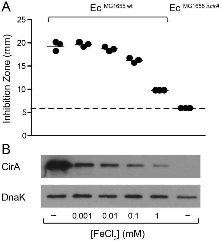
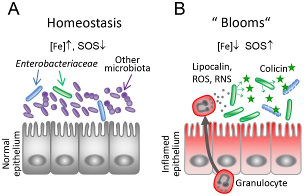
The host's immune system plays a key role in modulating growth of pathogens and the intestinal microbiota in the gut. In particular, inflammatory bowel disorders and pathogen infections induce shifts of the resident commensal microbiota which can result in overgrowth of Enterobacteriaceae ("inflammation-inflicted blooms"). Here, we investigated competition of the human pathogenic Salmonella enterica serovar Typhimurium strain SL1344 (S. Tm) and commensal E. coli in inflammation-inflicted blooms. S. Tm produces colicin Ib (ColIb), which is a narrow-spectrum protein toxin active against related Enterobacteriaceae. Production of ColIb conferred a competitive advantage to S. Tm over sensitive E. coli strains in the inflamed gut. In contrast, an avirulent S. Tm mutant strain defective in triggering gut inflammation did not benefit from ColIb. Expression of ColIb (cib) is regulated by iron limitation and the SOS response. CirA, the cognate outer membrane receptor of ColIb on colicin-sensitive E. coli, is induced upon iron limitation. We demonstrate that growth in inflammation-induced blooms favours expression of both S. Tm ColIb and the receptor CirA, thereby fuelling ColIb dependent competition of S. Tm and commensal E. coli in the gut. In conclusion, this study uncovers a so-far unappreciated role of inflammation-inflicted blooms as an environment favouring ColIb-dependent competition of pathogenic and commensal representatives of the Enterobacteriaceae family.


Conflict of interest statement

The authors declare no competing financial interests.

Figures

Figure 1
Figure 1. Colicin-dependent competition of S. Tm and E. coli in the gut in inflammation-induced “blooms” in gnotobiotic LCM mice.
Streptomycin-treated LCM-mice were co-infected with 1∶1 mixtures of S. TmΔoriT and EcMG1655, S. TmoriT Δcib and EcMG1655 or S. TmΔoriT avir and EcMG1655. (A) S. Tm (black) and E. coli (white) colonization densities were determined at day 4 p.i. in the cecum content (cfu/g). (B) Competitive indices (CI; ratio of S. Tm/E. coli) were determined for individual mice shown in (A). Bars show the median. (C) Histopathological analysis of cecal tissue of the infected mice shown in (A). Cecal tissue sections of the mice were stained with hematoxylin/eosin (H&E) and the degree of submucosal edema, neutrophil infiltration, epithelial damage and loss of goblet cells was scored (Materials and Methods). 1–3: no pathological changes; 4–7: moderate inflammation; above 8: severe inflammation. shown mean and StD. (D) Representative H&E–stained cecal sections of mice shown in (A–C). Magnification 100-fold. Enlarged sections (squares) are shown in the lower panels. Dotted line: detection limit (1 cfu/g).
Figure 2
Figure 2. Colicin-dependent competition of S. Tm and E. coli in the gut in inflammation-induced “blooms” in conventional mice.
Streptomycin-treated conventional mice were co-infected with 1∶1 mixtures of S. TmΔoriT Δcib and EcNissle, S. TmΔoriT and EcNissle, S. TmΔP2 avir and EcNissle or S. TmΔoriT avir and EcNissle. S. Tm (black) and E. coli (white) colonization densities (Cfu/g) were determined at day 1 (A) and day 4 p.i. (B) in the feces and cecum content respectively. (C, D) Competitive indices (CI; ratio of S. Tm/E. coli) were determined for individual mice shown in (A) and (B). Bars show the median. (E) Histopathological analysis of cecal tissue of the infected mice. Cecal tissue sections of the mice were stained with hematoxylin/eosin (H&E) and the degree of submucosal edema, neutrophil infiltration, epithelial damage and loss of goblet cells was scored (Materials and Methods). 1–3: no pathological changes; 4–7: moderate inflammation; above 8: severe inflammation. Bars show mean and StD. (F) Representative H&E–stained cecal sections of mice shown in (A–E). Magnification 100-fold. Enlarged sections (squares) are shown in the lower panels. Dotted line: detection limit (1 cfu/g).
Figure 3
Figure 3. Expression of S. Tm ColIb is induced by iron limitation and the SOS response.
(A) Organization of the ColIb locus showing location of the Fur and LexA repressor binding sites in the ColIb (cib) promoter and the immunity protein gene (imm). Overnight cultures of S. TmΔoriT (B) and S. TmΔoriT avir (C) carrying the reporter plasmid pcib -luc were re-inoculated 1∶20 in fresh LB with indicated supplements (0.25 µg/ml mitomycin C; 100 mM DTPA) and grown under aeration for 4 h. Cultures were normalized to OD600, bacteria were harvested and luciferase-activity was measured in bacterial lysates. Relative luminescence units (RLU) per unit OD600 are indicated. (D) Overnight cultures of indicated S. Tm strains were re-inoculated 1∶20 in fresh LB with indicated supplements (0.25 µg/ml mitomycin C; 100 mM DTPA) and grown under aeration for 4 h. OD600 of the cultures was normalized, bacteria were harvested and ColIb was detected in bacterial lysates as well as in the culture supernatant by immunoblot using an affinity-purified rabbit-α-ColIb antiserum. S. Tm DnaK was detected as loading control.
Figure 4
Figure 4. E. coli cirA expression is upregulated in response to iron limitation.
(A) An overnight culture of EcMG1655 carrying the reporter plasmid pcirA -luc was re-inoculated 1∶20 in fresh LB with indicated supplements (0.25 µg/ml mitomycin C; 100 mM DTPA) and grown under aeration for 4 h. OD600 of the cultures was normalized, bacteria were harvested and luciferase-activity was measured in bacterial lysates. Relative luminescence units (RLU) per unit OD600 are indicated. (B) Overnight cultures of EcMG1655 as well as EcMG1655 ΔcirA were re-inoculated 1∶20 in fresh LB with indicated supplements (0.25 µg/ml mitomycin C; 100 mM DTPA) and grown under aeration for 4 h. Cultures were normalized to OD600, bacteria were harvested and CirA was detected in bacterial lysates by immunoblot using a rabbit-α-CirA antiserum. E. coli cytoplasmic protein DnaK was detected as loading control.
Figure 5
Figure 5. Induction of cirA expression increases sensitivity to ColIb of E. coli.
(A) EcMG1655 and EcMG1655 ΔcirA were cultivated in M9 medium o.n., mixed with soft agar and indicated concentrations of FeCl3 and overlaid on M9 agar plates. Paper discs with recombinant ColIb were placed on the agar plate and the diameter of the inhibition zone (halo) was measured after 24 h. The detection limit (dotted line) is the size of the paper-disc (6 mm). (B) cirA expression of E. coli grown in vitro in M9 medium supplemented with different concentrations of FeCl3. Overnight cultures of EcMG1655 and EcMG1655 ΔcirA grown in M9 medium for 12 h were used for inoculation of 2 ml M9 medium supplemented with 1 µM, 10 µM, 0.1 mM or 1 mM FeCl3 and subcultured for 7 h. From each subculture, 250 µl (for an OD600 of 1) was taken, spun down at 4°C, 10 min, 10, 000 rpm. CirA was detected by immunoblot in bacterial lysates using affinity-purified rabbit-α-CirA-antiserum. E. coli DnaK was detected as loading control.
Figure 6
Figure 6. ColIb dependent competition of S. Tm and E. coli in vitro.
Overnight cultures of S. TmΔoriT and EcMG1655 (A, D, G, J), S. TmΔoriT Δcib and EcMG1655 (B, E, H, K) or S. TmΔoriT and EcMG1655 ΔcirA (C, F, I, L) were diluted and normalized to an OD600 of 0.05 for each strain in fresh LB with indicated supplements (0.25 µg/ml mitomycin C (Mito. C); 100 mM DTPA). Cfu/ml of both strains were determined at 0 h, 4 h, and 8 h after the start of the subculture. Red lines: S. Tm strains, blue lines: E. coli strains. Dotted line: detection limit (2000 cfu/ml).
Figure 7
Figure 7. S. Tm ColIb and the E. coli ColIb receptor CirA are induced in the inflamed gut.
To measure in vivo regulation of E. coli cirA expression, streptomycin-treated LCM-mice were co-infected with 1∶1 mixtures of the luciferase-reporter strain EcMG1655 pcirA -luc and S. TmΔoriT avir (avirulent) or S. TmΔoriT (wildtype), respectively (A, C). To measure in vivo regulation of S. Tm cib expression, streptomycin-treated LCM-mice were co-infected with the luciferase-reporter strain S. TmΔoriT pcib -luc and S. Tmavir (avirulent) or with S. Tmwt (wildtype), respectively (B, D). Bacteria were harvested from cecal content and luciferase-activity was measured in cecum content (A, B). Relative luminescence units (RLU) per 108 cfu of the reporter strain are indicated (C, D). Gut inflammation as determined by pathological score of cecal tissue sections of the infected mice. 1–3: no pathological changes; 4–7: moderate inflammation; above 8: severe inflammation. Bars show the median. Arrow in (A) and (C) point at one animal with atypically mild inflammatory symptoms.
Figure 8
Figure 8. Model for the role of colicins for bacterial competition in inflammation-induced blooms.
(A) Under homeostatic conditions, Enterobacteriaceae (blue, green) are reduced in numbers as they are kept in check by the obligate anaerobic microbiota (violet-blue). Under this condition, colicin expression as well as expression of the colicin surface receptors are relatively repressed (high iron, no triggers of the SOS response). (B) Upon induction of an inflammatory response, gut microbial ecology is altered leading to Enterobacterial blooms. Neutrophils transmigrate into the gut lumen and produce iron-depleting agents (lipocalin, lactoferrin) and reactive oxygen and nitrogen species (ROS, RNS). This triggers SOS-and Fur-dependent transcriptional responses in Enterobacteriaceae and colicin- and colicin-receptor expression is induced. Thereby, the inflammatory response drives colicin-dependent enterobacterial competition.

References

    1. Mushin R, Dubos R (1965) Colonization of the mouse intestine with Escherichia coli. J Exp Med 122: 745–757. - PMC - PubMed
    1. Barthel M, Hapfelmeier S, Quintanilla-Martinez L, Kremer M, Rohde M, et al. (2003) Pretreatment of mice with streptomycin provides a Salmonella enterica serovar Typhimurium colitis model that allows analysis of both pathogen and host. Infect Immun 71: 2839–2858. - PMC - PubMed
    1. Stecher B, Hardt WD (2010) Mechanisms controlling pathogen colonization of the gut. Curr Opin Microbiol 14 1: 82–91. - PubMed
    1. Sansonetti PJ (2008) Host-bacteria homeostasis in the healthy and inflamed gut. Curr Opin Gastroenterol 24: 435–439. - PubMed
    1. Lupp C, Robertson ML, Wickham ME, Sekirov I, Champion OL, et al. (2007) Host-mediated inflammation disrupts the intestinal microbiota and promotes the overgrowth of enterobacteriaceae. Cell, host and microbe 2: 119–129. - PubMed

Publication types