. 2014 Jan;10(1):e1003848.
doi: 10.1371/journal.ppat.1003848. Epub 2014 Jan 2.

DAMP molecule S100A9 acts as a molecular pattern to enhance inflammation during influenza A virus infection: role of DDX21-TRIF-TLR4-MyD88 pathway

Affiliations

DAMP molecule S100A9 acts as a molecular pattern to enhance inflammation during influenza A virus infection: role of DDX21-TRIF-TLR4-MyD88 pathway

Su-Yu Tsai et al. PLoS Pathog. 2014 Jan.

Abstract

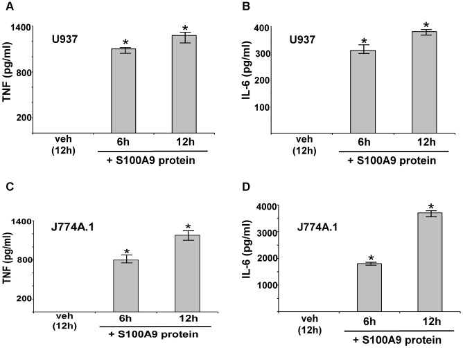
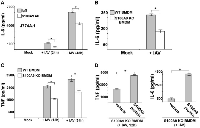
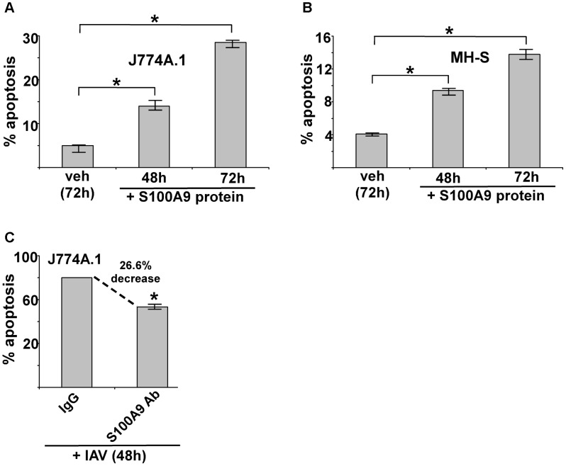
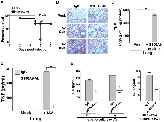
Pathogen-associated molecular patterns (PAMPs) trigger host immune response by activating pattern recognition receptors like toll-like receptors (TLRs). However, the mechanism whereby several pathogens, including viruses, activate TLRs via a non-PAMP mechanism is unclear. Endogenous "inflammatory mediators" called damage-associated molecular patterns (DAMPs) have been implicated in regulating immune response and inflammation. However, the role of DAMPs in inflammation/immunity during virus infection has not been studied. We have identified a DAMP molecule, S100A9 (also known as Calgranulin B or MRP-14), as an endogenous non-PAMP activator of TLR signaling during influenza A virus (IAV) infection. S100A9 was released from undamaged IAV-infected cells and extracellular S100A9 acted as a critical host-derived molecular pattern to regulate inflammatory response outcome and disease during infection by exaggerating pro-inflammatory response, cell-death and virus pathogenesis. Genetic studies showed that the DDX21-TRIF signaling pathway is required for S100A9 gene expression/production during infection. Furthermore, the inflammatory activity of extracellular S100A9 was mediated by activation of the TLR4-MyD88 pathway. Our studies have thus, underscored the role of a DAMP molecule (i.e. extracellular S100A9) in regulating virus-associated inflammation and uncovered a previously unknown function of the DDX21-TRIF-S100A9-TLR4-MyD88 signaling network in regulating inflammation during infection.


Conflict of interest statement

The authors have declared that no competing interests exist.

Figures

Figure 1
Figure 1. Production of S100A9 from IAV infected macrophages.
U937 cells (A), J774A.1 cells (B), primary bone marrow derived macrophages or BMDM (C) and primary mouse alveolar macrophages (D) were infected with IAV. U937 cells were infected at 1 MOI, whereas J774A.1, BMDM and primary alvelolar macrophages were infected at 2 MOI. At indicated post-infection time-periods the medium supernatant was collected to assess levels of S100A9 protein by ELISA. The values shown represent the mean ± standard deviation from three independent experiments performed in triplicate. *p<0.05 using a Student's t test.
Figure 2
Figure 2. DDX21 and TRIF is required for S100A9 production from IAV infected macrophages.
(A) Primary bone marrow derived macrophages or BMDMs isolated from wild-type (WT), TLR2 knockout (KO), TLR4 KO and TRIF KO mice were infected with IAV (2 MOI). At 24 h post-infection time-period the medium supernatant was collected to assess levels of S100A9 protein by ELISA. (B) RT-PCR analysis of S100A9 expression in IAV infected WT and TRIF KO BMDMs. (C) BMDMs isolated from WT and TLR3 KO mice were infected with IAV (2 MOI). At indicated post-infection time-periods the medium supernatant was collected to assess levels of S100A9 protein by ELISA. (D) Mouse alveolar macrophage cell-line MH-S was transfected with either control siRNA or DDX21 specific siRNA. At 48 h post-transfection, cells were infected with IAV (2 MOI). At indicated post-infection time-period RT-PCR analysis was performed to examine expression of DDX21 in IAV infected control and DDX21 silenced cells. (E) MH-S cells transfected with either control siRNA or DDX21 specific siRNA were infected with IAV (2 MOI). At indicated post-infection time-period the medium supernatant was collected to assess levels of S100A9 protein by ELISA. (F) BMDMs isolated from WT and TLR7 KO mice were infected with IAV (2 MOI). At indicated post-infection time-periods the medium supernatant was collected to assess levels of S100A9 protein by ELISA. The values shown in (A), (C), (E) and (F) represent the mean ± standard deviation from three independent experiments performed in triplicate. *p<0.05 using a Student's t test. Each RT-PCR data (B and D) is a representative of three independent experiments with similar results.
Figure 3
Figure 3. Extracellular S100A9 protein stimulates pro-inflammatory response in macrophages.
Human U937 macrophages were incubated with purified recombinant human S100A9 protein (10 µg/ml) for 6 h and 12 h. The medium supernatant was collected to assess levels of human TNF-α (TNF) (A) and human IL-6 (B) by ELISA. Mouse J774A.1 macrophages were incubated with purified recombinant mouse S100A9 protein (5 µg/ml) for 6 h and 12 h. The medium supernatant was collected to assess levels of mouse TNF (C) and mouse IL-6 (D) by ELISA. The values represent the mean ± standard deviation from three independent experiments performed in triplicate. *p<0.05 using a Student's t test. Vehicle control cells (veh) were incubated with HBSS buffer.
Figure 4
Figure 4. Extracellular S100A9 plays an essential role in inducing pro-inflammatory response during IAV infection of macrophages.
(A) Mouse J774A.1 macrophages were infected with IAV (2 MOI) in the presence of either control IgG (IgG) or anti-S100A9 blocking (neutralizing) antibody (S100A9 Ab). At indicated post-infection time-periods the medium supernatant was collected to assess levels of mouse IL-6 by ELISA. (B) Primary bone marrow derived macrophages (BMDM) isolated from wild-type (WT) and S100A9 knockout (KO) mice were infected with IAV (2 MOI). The medium supernatant was collected to assess levels of mouse IL-6 by ELISA. (C) WT and S100A9 KO BMDM were infected with IAV (2 MOI). At the indicated post-infection time-period, medium supernatant was collected to assess levels of mouse TNF-α(TNF) by ELISA. (D) S100A9 KO BMDMs were infected with IAV (2 MOI) in the presence of purified recombinant mouse S100A9 protein (5 µg/ml). Medium supernatant was collected from infected cells to assess levels of mouse TNF and IL-6 by ELISA. Vehicle control cells (veh) were incubated with HBSS buffer. The values represent the mean ± standard deviation from three independent experiments performed in triplicate. *p<0.05 using a Student's t test.
Figure 5
Figure 5. Extracellular S100A9 protein triggers apoptosis in macrophages and S100A9 regulates apoptosis during IAV infection.
(A) Mouse J774A.1 macrophages were incubated with purified recombinant mouse S100A9 protein (5 µg/ml) for 48 h and 72 h. The apoptotic state of these cells was examined by FACS analysis of annexin V and PI stained cells. Apoptosis rate (% apoptosis) was calculated based on number of annexin V positive/PI negative cells (denoting early apoptosis)+number of annexin V positive/PI positive cells (denoting late apoptosis)/total number of cells. (B) Mouse alveolar macrophage MH-S cell-line was incubated with purified S100A9 protein (5 µg/ml) for 48 h and 72 h. The apoptotic status was determined as described in (A). (C) Mouse J774A.1 macrophages were infected with IAV (2 MOI) in the presence of either control IgG (IgG) or anti-S100A9 blocking (neutralizing) antibody (S100A9 Ab). At 48 h post-infection, the apoptotic state of these cells was determined as described in (A). The values (i.e. annexin V and PI staining quantified by FACS) represents mean ± standard deviation from three independent experiments, *p<0.05 by Student's t test. Veh; cells incubated with HBSS buffer (vehicle control).
Figure 6
Figure 6. S100A9 activates TLR4/MyD88 pathway and activation of TLR4/MyD88 pathway is essential for IAV-induced pro-inflammatory response.
Primary bone marrow derived macrophages (BMDM) isolated from wild-type (WT) and TLR4 knockout (KO) mice were incubated with purified recombinant mouse S100A9 protein (5 ug/mL). The medium supernatant was collected to assess levels of mouse IL-6 (A) and mouse TNF-α(TNF) (B) by ELISA. (C) IL-6 production from S100A9 protein treated WT and MyD88 KO BMDMs. (D) BMDM isolated from WT, TLR4 KO and MyD88 KO mice were infected with IAV (2 MOI). At 12 h and 24 h post-infection time-period, medium supernatant was collected to assess levels of mouse IL-6 by ELISA. (E) TNF production from IAV infected WT and TLR4 KO BMDMs. The values represent the mean ± standard deviation from three independent experiments performed in triplicate, *p<0.05 using a Student's t test. Veh; cells incubated with HBSS buffer (vehicle control).
Figure 7
Figure 7. Activated TLR4/MyD88 pathway promotes S100A9-mediated apoptosis and is required for optimal apoptosis of IAV infected cells.
(A) Primary bone marrow derived macrophages (BMDM) isolated from wild-type (WT) and TLR4 knockout (KO) mice were incubated with purified recombinant mouse S100A9 protein (5 ug/mL) for 72 h. The apoptotic state of these cells was examined by FACS analysis of annexin V and PI stained cells. Apoptosis rate (% apoptosis) was calculated based on number of annexin V positive/PI negative cells (denoting early apoptosis)+number of annexin V positive/PI positive cells (denoting late apoptosis)/total number of cells. (B) WT and TLR4 KO BMDMs were infected with IAV (1 MOI). At 48 h post-infection, the apoptotic status was determined as described in (A). (C) IAV infected WT and TLR4 KO cells were subjected to TUNEL assay. TUNEL positive cells were analyzed by image J software. Percent TUNEL positive cells denotes ratio of number of TUNEL positive cells/total number of cells. (D) WT and MyD88 KO BMDMs were infected with IAV (1 MOI). At 48 h post-infection, the apoptotic status was determined. The values represents mean ± standard deviation from three independent experiments, *p<0.05 by Student's t test. Veh; cells incubated with HBSS buffer (vehicle control).
Figure 8
Figure 8. S100A9 expression and production in the IAV infected respiratory tract.
(A) RNA isolated from mock infected and IAV infected (2×10⁴ pfu/mouse via intra-tracheal route) mice were subjected to RT-PCR analysis to examine expression of mouse S100A9. The RT-PCR data represents three mice/group (i.e. three mock mice, three mice infected with IAV for 3 d and three mice infected with IAV for 6 d). The RT-PCR data is a representative of three independent experiments with similar results. (B) Lung homogenate prepared from mock infected and IAV infected (2×10⁴ pfu/mouse via intra-tracheal route) mice were subjected to ELISA analysis to determine levels of mS100A9 protein in the lung. (C) Immuno-histochemical analysis of mouse lung tissue sections derived from mock infected and IAV infected mice were stained with mouse S100A9 antibody. Magnification, 200×. One representative example of a total of 3 mice analyzed per group in two independent experiments. (D) Broncho-alveolar lavage fluid (BALF) isolated from mock infected and IAV infected (2×10⁴ pfu/mouse via intra-tracheal route) mice were subjected to ELISA analysis to determine levels of S100A9 protein in BALF. The values shown in (B) and (D) represent the mean ± standard deviation from three independent experiments performed in triplicate. *p<0.05 using a Student's t test.
Figure 9
Figure 9. S100A9 contributes to enhanced susceptibility and inflammation during IAV infection and S100A9 regulates pro-inflammatory response in the respiratory tract of IAV infected mice.
(A) Survival of IAV infected (1×10⁵ pfu/mouse via intra-tracheal route) mice administered with either control IgG (IgG) or anti-S100A9 blocking (neutralizing) antibody (S100A9 Ab) (24 h prior to IAV inoculation, 2 mg of antibody/mouse administered via i.p route). The data represents values from two independent experiments performed with 5 mice/group for each experiment (total 10 mice/group from two experiments); *p = 0.03. (B) Hematoxylin and eosin (H&E) staining of lung sections from mock infected or IAV infected mice (3×10⁴ pfu/mouse via intra-tracheal route) administered with either control IgG (IgG) or S100A9 Ab (24 h prior to IAV inoculation, 2 mg of antibody/mouse was administered via i.p route). Magnification, ×10. (C) Mice were administered with purified recombinant mouse S100A9 protein (15 µg/mouse) via intra-tracheal route. At 8 h post-administration, levels of mouse TNF-α in the lung was assessed by performing ELISA analysis with lung homogenate. (D) Lung homogenate prepared from mock infected and IAV infected (2×10⁴ pfu/mouse via intra-tracheal route) mice administered with either control IgG (IgG) or anti-S100A9 blocking (neutralizing) antibody (S100A9 Ab) (24 h prior to IAV inoculation, 2 mg of antibody/mouse administered via i.p route) were subjected to ELISA analysis to determine levels of mouse TNF-α in the lung. (E) For ex-vivo experiment, broncho-alveolar lavage fluid (BALF) was collected (at 3 d post-infection) from IAV infected mice (2×10⁴ pfu/mouse via intra-tracheal route) administered with either control IgG (IgG) or anti-S100A9 blocking (neutralizing) antibody (S100A9 Ab) (24 h prior to IAV inoculation, 2 mg of antibody/mouse administered via i.p route). The BALF cells were isolated and plated in 48-well plate. After 2 h and 4 h, the medium supernatant was analyzed for mouse TNF-α (TNF) and mouse IL-6 by ELISA. Values shown in (C), (D) and (E) represent the mean ± standard deviation from three independent experiments performed in triplicate. *p<0.05 using a Student's t test. Veh; HBSS buffer diluted in PBS (vehicle control).
Figure 10
Figure 10. Extracellular S100A9 promotes optimal apoptosis in the lung of IAV infected mice.
(A) Lung sections were prepared (at 3 d post-infection) from IAV infected (2×10⁴ pfu/mouse via intra-tracheal route) mice administered with either control IgG (IgG) or anti-S100A9 blocking (neutralizing) antibody (S100A9 Ab) (24 h prior to IAV inoculation, 2 mg of antibody/mouse administered via i.p route). For each experimental group lung sections were prepared from three control IgG treated mice (+IAV) and three S100A9 Ab treated mice (+IAV). The lung sections were used for TUNEL staining. Image J software was used to calculate TUNEL-positive areas (representing apoptosis) in the lung sections as detailed in the methods section. The data is presented as percent apoptotic area. The percent apoptotic area was calculated from nine areas/lung section as detailed in the methods section. The values were compiled to calculate the percent apoptotic area in IAV infected IgG treated mice vs. IAV infected S100A9 Ab treated mice, *p = 0.0164 by Student's t test. (B) A representative TUNEL staining of lung sections from IAV infected mice administered with either IgG or S100A9 Ab. The apoptotic nuclei (representing apoptosis) are indicated with red arrows. (C) A schematic model depicting the role of extracellular S100A9 and DDX21/TRIF/S100A9/TLR4/MyD88 signaling network in exaggerating lung disease during IAV infection. PM, plasma membrane; NM, nuclear membrane.

Similar articles

Cited by

References

    1. Kawai T, Akira S (2010) The role of pattern-recognition receptors in innate immunity: Update on toll-like receptors. Nat Immunol 11: 373–384. - PubMed
    1. McGettrick AF, O'Neill LAJ (2010) Localisation and trafficking of toll-like receptors: An important mode of regulation. Curr Opin Immunol 22: 20–27. - PubMed
    1. Piccinini AM, Midwood KS (2010) DAMPening inflammation by modulating TLR signalling. Mediators Inflamm 2010: pii: 672395. - PMC - PubMed
    1. Kunisaki KM, Janoff EN (2009) Influenza in immunosuppressed populations: A review of infection frequency, morbidity, mortality, and vaccine responses. Lancet Infect Dis 9: 493–504. - PMC - PubMed
    1. Tscherne DM, Garcia-Sastre A (2011) Virulence determinants of pandemic influenza viruses. J Clin Invest 121: 6–13. - PMC - PubMed

Publication types

MeSH terms