. 2014 Jan;10(1):e1003854.
doi: 10.1371/journal.ppat.1003854. Epub 2014 Jan 2.

Recruitment of PfSET2 by RNA polymerase II to variant antigen encoding loci contributes to antigenic variation in P. falciparum

Affiliations

Recruitment of PfSET2 by RNA polymerase II to variant antigen encoding loci contributes to antigenic variation in P. falciparum

Uchechi E Ukaegbu et al. PLoS Pathog. 2014 Jan.

Abstract

Histone modifications are important regulators of gene expression in all eukaryotes. In Plasmodium falciparum, these epigenetic marks regulate expression of genes involved in several aspects of host-parasite interactions, including antigenic variation. While the identities and genomic positions of many histone modifications have now been cataloged, how they are targeted to defined genomic regions remains poorly understood. For example, how variant antigen encoding loci (var) are targeted for deposition of unique histone marks is a mystery that continues to perplex the field. Here we describe the recruitment of an ortholog of the histone modifier SET2 to var genes through direct interactions with the C-terminal domain (CTD) of RNA polymerase II. In higher eukaryotes, SET2 is a histone methyltransferase recruited by RNA pol II during mRNA transcription; however, the ortholog in P. falciparum (PfSET2) has an atypical architecture and its role in regulating transcription is unknown. Here we show that PfSET2 binds to the unphosphorylated form of the CTD, a property inconsistent with its recruitment during mRNA synthesis. Further, we show that H3K36me3, the epigenetic mark deposited by PfSET2, is enriched at both active and silent var gene loci, providing additional evidence that its recruitment is not associated with mRNA production. Over-expression of a dominant negative form of PfSET2 designed to disrupt binding to RNA pol II induced rapid var gene expression switching, confirming both the importance of PfSET2 in var gene regulation and a role for RNA pol II in its recruitment. RNA pol II is known to transcribe non-coding RNAs from both active and silent var genes, providing a possible mechanism by which it could recruit PfSET2 to var loci. This work unifies previous reports of histone modifications, the production of ncRNAs, and the promoter activity of var introns into a mechanism that contributes to antigenic variation by malaria parasites.


Conflict of interest statement

The authors have declared that no competing interests exist.

Figures

Figure 1
Figure 1. Two histone modifiers unique to primate parasites.
A comparative screen of the genomes of primate and rodent malaria parasites identified two histone modifiers unique to P. falciparum, P. vivax and P. knowlesi, SET2 and JmjC1. (A) Domain searches of the amino acid sequences using SMART (Simple Modular Architecture Research Tool, http://smart.embl-heidelberg.de) identified several domains typical of chromatin modifying enzymes, including several PHD domains and a SET2-like methyltransferase domain in PfSET2 and a predicted JmjC type demethylase in PfJmjC1. (B) Syntenic regions of chromosomes 13 and 8 of three primate parasites and three rodent parasites. Extensive synteny is observed throughout these chromosomal regions, with the exception of SET2 and JmjC1, which are missing from the genomes of the rodent parasites. The flanking genes are highly conserved. Figure adapted from Plasmodb.org.
Figure 2
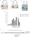
Figure 2. Detection of H3K36me3 in chromatin from P. falciparum and identification of the Rpb1binding region of PfSET2.
(A) Slot blots of the synthetic peptide sequence RKSAPISAGIKKPHRYRPGT. This sequence includes K36 of histone H3 of P. falciparum (underlined). Lane 1, unmethylated peptide. Lane 2, peptide synthesized with a mono-methyl group at the K36 position. Lane 3, peptide synthesized with a di-methyl group at the K36 position. Lane 4, peptide synthesized with a tri-methyl group at K36 position. All lanes included 15 µg of purified peptide. The dot blot was probed with antibodies against the tri-methylated form of the peptide and demonstrated that the antibodies are specific to the tri-methylated form. (B) Western blot of P. falciparum protein extracts taken from early trophozoites. Lane 1, blot of total parasite proteins probed with anti-H3K36me3 antibodies. Lane 2, blot of total parasite proteins probed with anti-H3-core antibodies. Both antibodies recognize bands migrating at ∼18 kDa. Lane 3, blot of acid extracted histones obtained from cultured parasites and probed with anti-H3K36me3 antibodies. (C) Comparison of the architecture of SET2 orthologs from S. cerevisiae, H. sapien and P. falciparum. Note that the SET2-Rpb1-Interacting domain (SRI) is found at the C-terminal end of both the yeast and human SET2 proteins. The protein domains shown in the figure are displayed using the format derived from SMART (Simple Modular Architecture Research Tool, http://smart.embl-heidelberg.de). Below PfSET2 are displayed the portions produced as HA-tagged, recombinant proteins for use in co-immunoprecipitation assays. The numbers below each fragment represent the amino acid positions within the full-length protein. The tandem PHD domains (amino acids 829–1060) were not included because the DNA encoding the this region of the protein was unstable in the expression vector. The SET domain was also not included because the structure and function of this domain is conserved and was previously shown not to bind the CTD in yeast and human systems. (D) Co-immunoprecipitation assays used to identify the region of PfSET2 that interacts with the CTD of Rpb1. Flag-tagged fragments of the CTD of Rpb1 shown in E were incubated with bacterial lysates containing the truncates of PfSET2 shown in C. Immunoprecipitation was performed using anti-HA beads followed by SDS-PAGE and Western blotting using anti-Flag antibodies. Only the most N-terminal portion of PfSET2 (amino acids 1–359) was found to interact with the CTD of Rpb1. The control reaction consisted of an HA-tagged PHIST protein similarly incubated with the CTD of Rpb1. (E) The N-terminal region of PfSET2 also binds to Rpb1 regions with fewer numbers of repeats. Co-immunoprecipitation assay performed as in D, using either the CTD of Rpb1 from P. falciparum (left lane) or P. berghei (right lane). The P. berghei CTD has only 8 heptad repeats.
Figure 3
Figure 3. Phosphorylation dependence of PfSET2 binding to the CTD and enrichment of H3K36me3 at var loci.
(A) Co-immunoprecipitation assays showing that phosphorylation of the CTD of Rpb1 eliminates binding by PfSET2. The two left lanes show the flag-tagged P. falciparum Rpb1 in its phosphorylated (IIo) or unphosphorylated (IIa) states. The right two lanes show blots of co-immunoprecipitation assays after incubation of bacterial lysates containing the HA tagged, N-terminal region of PfSET2 (PfSET21–359) with phosphorylated and unphosphorylated CTD. Phosphorylation of the CTD disrupts binding by PfSET2. (B) Co-immunoprecipitation assays as in A performed with the CTD of Rpb1 from P. berghei. (C) Direct comparison of the CTD binding affinities of SET2 from S. cerevisiae and P. falciparum. The SRI from S. cerevisiae (left two lanes) interacts preferentially with the phosphorylated form of the P. falciparum CTD (left) but also displays lesser binding to the unphosphorylated form (right). The right two lanes show the same assay with the P. falciparum SRIR region (amino acids 1–359). The SRIR displays specific binding to the unphosphorylated CTD (right) and little or no binding to the phosphorylated form (left).(D) Chromatin immunoprecipitation assays from ring stage parasites using anti-H3K36me3 antibodies. The assays show enrichment of the H3K36me3 mark in both exons of var gene PF3D7_0421100. Lanes 1 and 2 represent control genes encoding seryl t-RNA synthetase and actin, respectively. Lane 3 represents the gene for circumsporozoite protein. The remaining lanes represent regions within the coding portion of both exons of the var gene PF3D7_0421100. Black bars show results from chromatin extracted from the C3 line of NF54, in which PF3D7_0421100 is the actively expressed var gene. The gray bars show chromatin extracted from the A3 line of NF54 in which this var gene is transcriptionally silent. The bars display the mean +/− standard deviation of relative amounts of bound DNA (see methods) from four independent experiments. Similar experiments using no-specific antibodies or control antibodies against core histone H3 showed no specific enrichment at PF3D7_0421100 (not shown). The graph shown represents the signal for each gene normalized to the control gene ctrp. The data is shown as % input without normalization in Figure S7. (E) Schematic representation of a typical var gene. Note that each gene has three promoters, one upstream of the coding region and responsible for transcribing an mRNA from the single, active var gene and two within the intron transcribing ncRNAs in opposite directions. The intron promoter that transcribes the antisense ncRNA is active during rings stages and is only active in genes that are also transcribing mRNA. The intron promoter transcribing ncRNA in the sense direction leads to expression of “sterile”, ncRNAs during late trophozoites and schizonts and is active in most or all members of the var gene family.
Figure 4
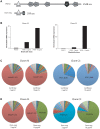
Figure 4. Over-expression of a dominant-negative version of PfSET2 in cultured parasites.
(A) Schematic diagrams showing the domain structures of the endogenous PfSET2 protein (top) and the truncated, dominant-negative version that contains only the N-terminal 359 amino acids (bottom), including the CTD-binding domain (SRIR). (B) Histograms showing RNA expression levels of the endogenous PfSET2 gene (PfSET2) and the dominant-negative construct (PfSRIR) under 2 and 10 µg/ml blasticidin. Two separate subclones of NF54 were transfected, A3 (left) and C3 (right). (C and D) Pie charts representing var gene expression patterns for A3 (left) and C3 (right) when over-expressing either firefly luciferase (C) or the PfSET2 dominant-negative construct (D). The different wedges of the pie indicate the proportion of the total var mRNA pool represented by transcripts for each individual gene (as described in [28]).

Similar articles

Cited by

References

    1. Jiang L, Lopez-Barragan MJ, Jiang H, Mu J, Gaur D, et al. (2010) Epigenetic control of the variable expression of a Plasmodium falciparum receptor protein for erythrocyte invasion. Proc Natl Acad Sci U S A 107: 2224–2229. - PMC - PubMed
    1. Cortes A, Carret C, Kaneko O, Yim Lim BY, Ivens A, et al. (2007) Epigenetic silencing of Plasmodium falciparum genes linked to erythrocyte invasion. PLoS Pathog 3: e107. - PMC - PubMed
    1. Crowley VM, Rovira-Graells N, Ribas dP, Cortes A (2011) Heterochromatin formation in bistable chromatin domains controls the epigenetic repression of clonally variant Plasmodium falciparum genes linked to erythrocyte invasion. Mol Microbiol 80: 391–406. - PubMed
    1. Comeaux CA, Coleman BI, Bei AK, Whitehurst N, Duraisingh MT (2011) Functional analysis of epigenetic regulation of tandem RhopH1/clag genes reveals a role in Plasmodium falciparum growth. Mol Microbiol 80: 378–390. - PMC - PubMed
    1. Chookajorn T, Dzikowski R, Frank M, Li F, Jiwani AZ, et al. (2007) Epigenetic memory at malaria virulence genes. Proc Natl Acad Sci U S A 104: 899–902. - PMC - PubMed

Publication types

MeSH terms

LinkOut - more resources