. 2014 Jan;10(1):e1004055.
doi: 10.1371/journal.pgen.1004055. Epub 2014 Jan 2.

The candidate splicing factor Sfswap regulates growth and patterning of inner ear sensory organs

Affiliations

The candidate splicing factor Sfswap regulates growth and patterning of inner ear sensory organs

Yalda Moayedi et al. PLoS Genet. 2014 Jan.

Abstract

The Notch signaling pathway is thought to regulate multiple stages of inner ear development. Mutations in the Notch signaling pathway cause disruptions in the number and arrangement of hair cells and supporting cells in sensory regions of the ear. In this study we identify an insertional mutation in the mouse Sfswap gene, a putative splicing factor, that results in mice with vestibular and cochlear defects that are consistent with disrupted Notch signaling. Homozygous Sfswap mutants display hyperactivity and circling behavior consistent with vestibular defects, and significantly impaired hearing. The cochlea of newborn Sfswap mutant mice shows a significant reduction in outer hair cells and supporting cells and ectopic inner hair cells. This phenotype most closely resembles that seen in hypomorphic alleles of the Notch ligand Jagged1 (Jag1). We show that Jag1; Sfswap compound mutants have inner ear defects that are more severe than expected from simple additive effects of the single mutants, indicating a genetic interaction between Sfswap and Jag1. In addition, expression of genes involved in Notch signaling in the inner ear are reduced in Sfswap mutants. There is increased interest in how splicing affects inner ear development and function. Our work is one of the first studies to suggest that a putative splicing factor has specific effects on Notch signaling pathway members and inner ear development.


Conflict of interest statement

The authors have declared that no competing interests exist.

Figures

Figure 1
Figure 1. Mutation of Sfswap results in mice with vestibular and cochlear defects.
(A): Lentiviral integration into the 4th intron of Sfswap. The exons of Sfswap are shown with black vertical bars. The features of the lentivirus are indicated as follows: LTR: long terminal repeat, Psi: packaging sequence, cPPT: central polypurine tract, Enh: tyrosinase enhancer, Pro: tyrosinase promoter, WPRE: woodchuck hepatitis virus post-transcriptional regulatory element. Features of the Sfswap gene are indicated as follows: C: Coil-Coil, S: SURP domain, RS: Arginine-Serine domain. (B): Northern blot of brain RNA from wild-type and SfswapTg mice. The mutation caused by the insertion of the lentivirus results in a reduction of wild-type Sfswap transcript (black arrow) and the appearance of a new isoform greater than 10 Kb in size (grey arrow). (C, D): In a 30-minute open field task, SfswapTg mice exhibit increased circling behavior (C) and hyperactivity (D). (E, F): SfswapTg/Tg mice exhibit increased auditory thresholds measured by ABR (E; p(WT, Tg/Tg) = 0.003, p(Tg/+, Tg/Tg) = 2×10⁻⁴) (black - WT, grey - Tg/+, red - Tg/Tg) and increased DPOAE thresholds (F; p(WT, Tg/Tg) = 1×10⁻⁴, p(Tg/+, Tg/Tg) = 3×10⁻⁶), indicating a hearing deficit. (G): SfswapTg/Tg mice show a reduced startle response. Mice were allowed to acclimate to a 70 dB background noise, and were then exposed to noise of increasing sound pressures. Wild type and Sfwap Tg/+ mice begin to show a response at 100 dB, whereas SfswapTg/Tg mice do not begin to show a startle response until 118 dB (p(WT, Tg/Tg) = 2.2×10⁻⁷, p(Tg/+, Tg/Tg) = 1.8×10⁻⁷).
Figure 2
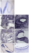
Figure 2. Sfswap is expressed in the developing inner ear.
(A): Sfswap is expressed in the E10.5 inner ear and hindbrain. (B, C): At E13.5, Sfswap RNA is expressed broadly throughout the wild-type cochlea and surrounding mesenchyme. The white box in (B) is shown at higher magnification in (C). (D, E): At P0, Sfswap expression is more restricted to the wild-type cochlea and the spiral ganglion within the inner ear and is expressed less strongly in surrounding tissues. Note that Sfswap is also expressed strongly in the hair follicles and dermis of neonatal mice (D). (F, G): Sfswap is expressed in the P0 cristae (F) and maculae of the utricle (F) and saccule (G).
Figure 3
Figure 3. Mutation of Sfswap results in a shorter cochlea, fewer outer hair cells and ectopic inner hair cells.
(A): Paint fills of E15.5 wild-type and SfswapTg/Tg inner ears. The main components of the inner ear labyrinth are all present, although the utricle (red arrow) and saccule (white arrow) appear smaller and the cochlea (yellow arrow) is reduced in length. (B): SfswapTg/Tg mice are missing hair cells in the third row of outer hair cells (dotted lines) in the basal and mid-turn regions of the cochlea. They also have extra inner hair cells near the apex (arrows). Scale bar = 20 µm. Phal: Phalloidin (C): The distribution of inner and outer hair cells is shown for the apical, mid-turn and basal thirds of the cochlea at P0. Hair cells were counted in 200 µm lengths. The change in hair cell numbers in SfswapTg/Tg mice is shown compared to wild-type controls. Significance is indicated as asterisks and given in Table 1 ** : p≤10−3, *** : p≤10−4, ****** : p≤10−7. (D): Total inner and outer hair cell counts for SfswapTg/Tg mice and wild-type controls (see also Table 1). The decrease in total cell counts reflects the decrease in the length of the mutant cochlea.
Figure 4
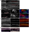
Figure 4. Mutation of Sfswap leads to fewer supporting cells.
(A): Cochleas from P0 SfswapTg/Tg and wild-type mice co-stained with Prox1 to show supporting cells and with fluorescently-labeled phalloidin (Phal) to reveal hair cells. In areas where hair cells are missing, supporting cells are also missing (white line compared to red line). Scale bar = 20 µm (B): Sections of P0 SfswapTg/Tg and wild-type mice co-stained with Prox1 (green) to show supporting cells and parvalbumin (Parv, red) to show hair cells. Missing hair cells are accompanied by missing supporting cells (asterisks). In addition, ectopic parvalbumin-expressing cells are observed in SfswapTg/Tg mutants (arrow). Scale bar = 10 µm (C, D): Assays for the pillar cell markers p75 (red) at P0 and β-tectorin (purple) at P2 shows occasional loss of pillar cells in SfswapTg/Tg mutants. Scale bar = 20 µm.
Figure 5
Figure 5. Mutation of Sfswap causes smaller vestibular cristae and maculae and misplaced eminentia cruciata.
(A, B): Flat mount preparations of utricles and saccules and cristae from P0 SfswapTg/Tg and wild-type mice stained with fluorescently-labeled phalloidin to reveal hair cells. In addition to reductions in size (C), anterior semicircular canal eminentia cruciata are reduced in 85% of ears examined (B, red arrows denote eminentia cruciata). U: Utricle, S: Saccule, AC: Anterior crista, PC: Posterior crista, HC: Horizontal crista. Scale bars = 50 µm. Significant p-values are denoted with asterisks ** : p≤10−3, *** : p≤10−4, **** : p≤10−5 (D): Sensory patch primordia are reduced in size as revealed by in situ hybridization for Bmp4 and Lfng.
Figure 6
Figure 6. Sfswap interacts genetically with Jagged1.
(A, B): Paint fills of E15.5 wild-type, SfswapTg/Tg, compound mutant (SfswapTg/Tg, Jag1+/−), and Jag1+/− inner ears. In the mixed FVBN/C57BL6 F2 background, semicircular canal truncations are not observed in SfswapTg/Tg or Jag1+/− mice. However, in SfswapTg/Tg, Jag1+/− mice, canal truncations are observed in 10/14 ears (arrow). Canal truncations are observed in Jag1−/+ mutants on the C57BL6 background (B). However, when crossed one generation to FVB, this phenotype is suppressed (B, F1). The numbers in each panel refer to the number of ears with canal truncations. (C): An enhancement of hair cell phenotypes is observed in SfswapTg/Tg, Jag1+/− mutants compared to either SfswapTg/Tg or Jag1+/− mice. These enhanced defects include more ectopic inner hair cells in the base and mid-turn regions (arrowheads and brackets), loss of second and third rows of outer hair cells (white dashed line), and a 4th row of outer hair cells in the apex (red dashed line). Scale bars = 20 µm. (D): The length of the SfswapTg/Tg, Jag1+/− mutant cochlea is significantly shorter than wild-type, SfswapTg/Tg or Jag1+/− mice. Asterisks denote p-values defined in Table 2 (* : p≤0.05, ***** : p≤10−6). (E): SfswapTg/Tg, Jag1+/− mutants have significantly fewer third row outer hair cells than either Jag1−/+ or SfswapTg/Tg mice in the mid-turn and basal regions. Hair cells were counted in 200 µm lengths, and the change in hair cell numbers in SfswapTg/Tg, SfswapTg/Tg, Jag1+/− or Jag1+/− cochleas is shown compared to wild-type controls. (F) Jag1+/− mutants have significantly increased ABR thresholds particularly at high frequencies. Furthermore, SfswapTg/Tg, Jag1+/− mutant ABR thresholds are significantly increased compared to either SfswapTg/Tg or Jag+/− mutants. (G) Jag1+/− mutants have increased DPOAE thresholds at high frequencies compared to WT. SfswapTg/Tg, Jag1+/− have increased DPOAE thresholds compared to either SfswapTg/Tg or Jag+/− mutants.
Figure 7
Figure 7. Jagged1 expression is not altered in SfswapTg/Tg ears but its downstream target Hey1 is reduced at E10.5.
(A): Cochleas from E13.5 SfswapTg/Tg and wild-type mice co-stained with antibodies to p27kip1 (green) to reveal the prosensory domain and Jag1 (red) to show its expression in the adjacent Kölliker's organ. No significant difference is seen in Jag1 expression in the mutant cochlea. (B): Whole mount in situ hybridization of E10.5 SfswapTg/Tg and wild-type mice reveals no significant change in Jag1 expression, but a significant down-regulation of its candidate downstream target, Hey1 (C). Otocysts are denoted with white arrow. (D): cDNAs from E15.5 SfswapTg/Tg and wild-type inner ears were examined for splicing differences in Notch pathway genes by RT-PCR. No significant differences in splicing were detected in any gene except for Sfswap. The extra band in Sfswap mRNA in SfswapTg/Tg corresponds to 111 bp of transgene sequence that is spliced into the Sfswap mRNA exclusively in homozygous SfswapTg mice (data not shown). Levels of Neurl1 Numb mRNA were reduced, and MamlD1 expression was not detected. X refers to no cDNA controls.

Similar articles

Cited by

References

    1. Lewis AK, Frantz GD, Carpenter DA, de Sauvage FJ, Gao WQ (1998) Distinct expression patterns of notch family receptors and ligands during development of the mammalian inner ear. Mech Dev 78: 159–163. - PubMed
    1. Murata J, Tokunaga A, Okano H, Kubo T (2006) Mapping of notch activation during cochlear development in mice: implications for determination of prosensory domain and cell fate diversification. J Comp Neurol 497: 502–518. - PubMed
    1. Morrison A, Hodgetts C, Gossler A, Hrabe de Angelis M, Lewis J (1999) Expression of Delta1 and Serrate1 (Jagged1) in the mouse inner ear. Mech Dev 84: 169–172. - PubMed
    1. Hartman BH, Hayashi T, Nelson BR, Bermingham-McDonogh O, Reh TA (2007) Dll3 is expressed in developing hair cells in the mammalian cochlea. Dev Dyn 236: 2875–2883. - PubMed
    1. Kiernan AE, Cordes R, Kopan R, Gossler A, Gridley T (2005) The Notch ligands DLL1 and JAG2 act synergistically to regulate hair cell development in the mammalian inner ear. Development 132: 4353–4362. - PubMed

Publication types

MeSH terms