Review
. 2013 Dec 19:7:256.
doi: 10.3389/fncel.2013.00256.

Concepts for regulation of axon integrity by enwrapping glia

Affiliations
Review

Concepts for regulation of axon integrity by enwrapping glia

Bogdan Beirowski. Front Cell Neurosci. .

Abstract

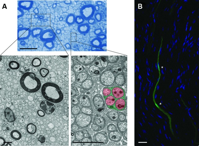
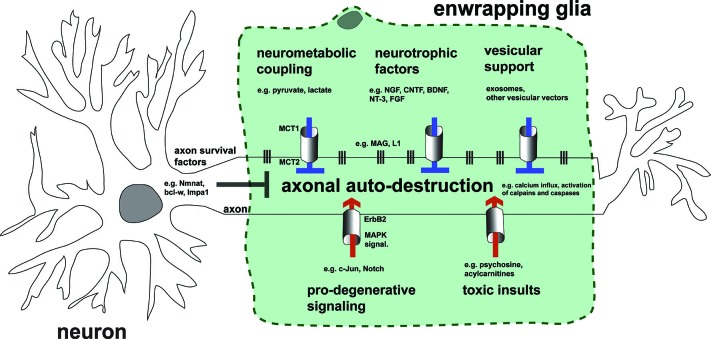
Long axons and their enwrapping glia (EG; Schwann cells (SCs) and oligodendrocytes (OLGs)) form a unique compound structure that serves as conduit for transport of electric and chemical information in the nervous system. The peculiar cytoarchitecture over an enormous length as well as its substantial energetic requirements make this conduit particularly susceptible to detrimental alterations. Degeneration of long axons independent of neuronal cell bodies is observed comparatively early in a range of neurodegenerative conditions as a consequence of abnormalities in SCs and OLGs . This leads to the most relevant disease symptoms and highlights the critical role that these glia have for axon integrity, but the underlying mechanisms remain elusive. The quest to understand why and how axons degenerate is now a crucial frontier in disease-oriented research. This challenge is most likely to lead to significant progress if the inextricable link between axons and their flanking glia in pathological situations is recognized. In this review I compile recent advances in our understanding of the molecular programs governing axon degeneration, and mechanisms of EG's non-cell autonomous impact on axon-integrity. A particular focus is placed on emerging evidence suggesting that EG nurture long axons by virtue of their intimate association, release of trophic substances, and neurometabolic coupling. The correction of defects in these functions has the potential to stabilize axons in a variety of neuronal diseases in the peripheral nervous system and central nervous system (PNS and CNS).

Keywords: Charcot-Marie-Tooth disease; amyotrophic lateral sclerosis; axon; multiple sclerosis; neurodegeneration; oligodendrocyte; schwann cell; wallerian degeneration.


Figures

Figure 1
Figure 1
Cytoarchitecture of EG and associated axons in mouse nerves. (A) (Upper): Semithin microscopy of transverse section through mouse vagus nerve in which more than 90% of all fibers are unmyelinated and form multiple Remak bundles. Scale bar: 10 µm. Boxed areas from semithin preparation show electron micrographs (Lower) at different magnifications with characteristic cytoarchitecture of unmyelinating and myelinating SCs with their enwrapped axons. Note the tight association between glial processes (green) that interdigitate four unmyelinated small-caliber axons (red) in the highlighted Remak bundle from the right inset. Individual Remak bundles are surrounded by basement membranes and collagen fibers. Axonal and glial mitochondria are characterized by electron-dense appearance. Granular material within axons represents microtubules and neurofilaments. Scale bars: 2 µm. (B) Fluorescence confocal microscopy of longitudinal section through mouse tibial nerve (nuclear counterstain with 4’,6-Diamidino-2-Phenylindole (DAPI)) in which two adjacent SC bodies (green) are genetically labeled with green fluorescent protein (GFP). Asterisks depict nuclear regions of SCs with their elongated bodies. Scale bar: 5 µm.
Figure 2
Figure 2
Schematic illustration showing scaled model of human neuron/axon (black) in association with approximately 10,000 SCs (green) in proportion to size of neuronal soma and axon terminal. Note that axons in humans or larger mammals often traverse distances of 1 m or longer and are accompanied by SCs almost over their full length. Inset shows higher magnification of boxed area in scaled model.
Figure 3
Figure 3
Hypothetical model summarizing impact of EG on axonal integrity by the mechanistic themes discussed. Preservation of healthy axons is controlled by the cooperative action of both the neuron and adjacent glia by blocking endogenous axonal auto-destruction. Examples of neuronal mechanisms blocking this program(s) include somatic delivery of the putative axonal survival molecule Nmnat2 into axons, or the local translation of axonal maintenance factors such as Bcl-w. On the other hand, glia inhibit axonal death by transfer of metabolic substrates, neurotrophic factors, and vesicular shuttles (upper glia portion). This transfer is mediated by specific transport mechanisms represented by columns between glia and axon. Vertical lines embody adhesion mechanisms that ensure correct apposition and formation of nutritive channels between glia and axon. Under pathological conditions glia may also contribute to axonal auto-destruction by activating pro-degenerative signaling, releasing toxic substances, and loosening contact to axons (lower glia portion). Note that the myelin membrane of EG is uncoiled in the illustration and myelination thus not represented.

References

    1. Adriano E., Perasso L., Panfoli I., Ravera S., Gandolfo C., Mancardi G., et al. (2011). A novel hypothesis about mechanisms affecting conduction velocity of central myelinated fibers. Neurochem. Res. 36, 1732–1739 10.1007/s11064-011-0488-0 - DOI - PubMed
    1. Agthong S., Kaewsema A., Tanomsridejchai N., Chentanez V. (2006). Activation of MAPK ERK in peripheral nerve after injury. BMC Neurosci. 7:45 10.1186/1471-2202-7-45 - DOI - PMC - PubMed
    1. Aguayo A. J., Bray G. M., Perkins S. C. (1979). Axon-Schwann cell relationships in neuropathies of mutant mice. Ann. N Y Acad. Sci. 317, 512–531 10.1111/j.1749-6632.1979.tb56571.x - DOI - PubMed
    1. Ahmed Z., Doward A. I., Pryce G., Taylor D. L., Pocock J. M., Leonard J. P., et al. (2002). A role for caspase-1 and -3 in the pathology of experimental allergic encephalomyelitis : inflammation versus degeneration. Am. J. Pathol. 161, 1577–1586 10.1016/s0002-9440(10)64436-7 - DOI - PMC - PubMed
    1. Ali Y. O., Li-Kroeger D., Bellen H. J., Zhai R. G., Lu H. C. (2013). NMNATs, evolutionarily conserved neuronal maintenance factors. Trends Neurosci. 36, 632–640 10.1016/j.tins.2013.07.002 - DOI - PMC - PubMed

LinkOut - more resources