. 2013 Dec 18:7:73.
doi: 10.3389/fnsys.2013.00073. eCollection 2013.

High and low frequency stimulation of the subthalamic nucleus induce prolonged changes in subthalamic and globus pallidus neurons

Affiliations

High and low frequency stimulation of the subthalamic nucleus induce prolonged changes in subthalamic and globus pallidus neurons

Hagar Lavian et al. Front Syst Neurosci. .

Abstract

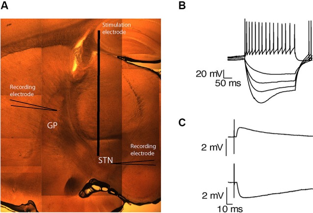
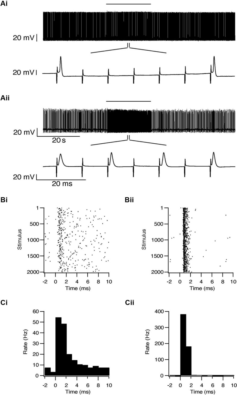
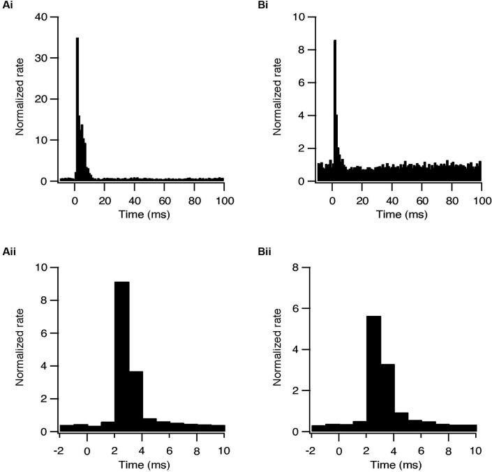
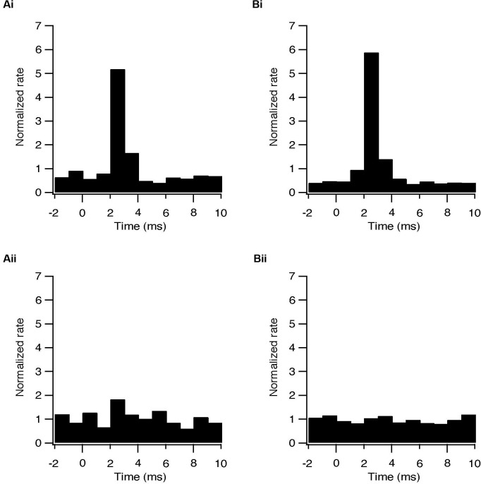
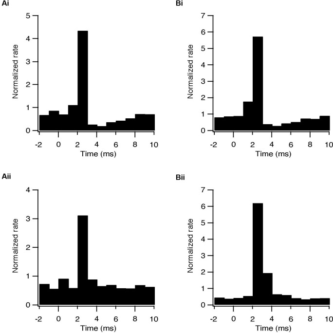
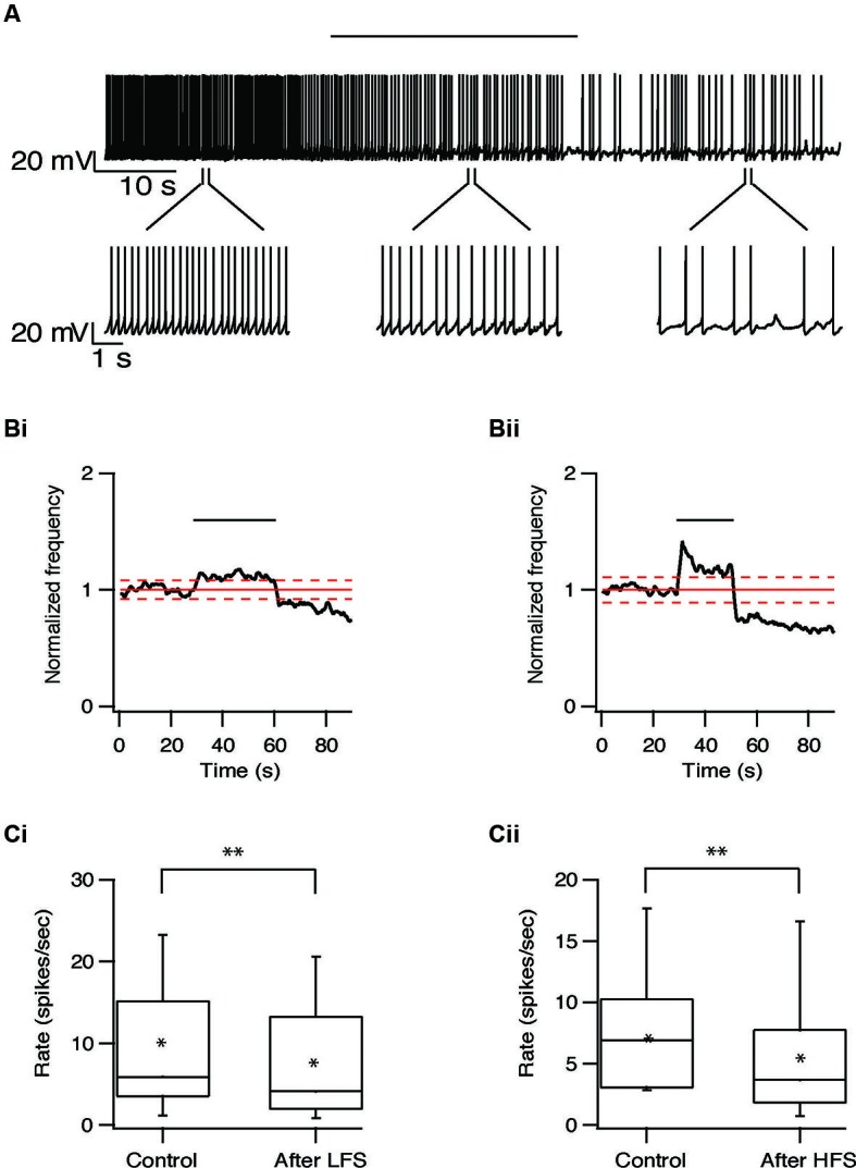
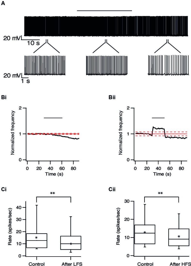
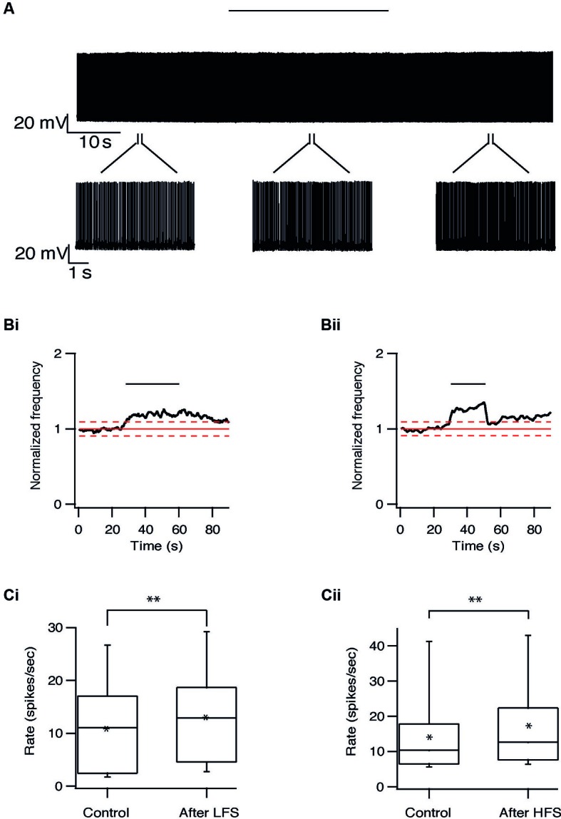
High frequency stimulation (HFS) of the subthalamic nucleus (STN) is widely used to treat the symptoms of Parkinson's disease (PD) but the mechanism of this therapy is unclear. Using a rat brain slice preparation maintaining the connectivity between the STN and one of its target nuclei, the globus pallidus (GP), we investigated the effects of high and low frequency stimulation (LFS) (HFS 100 Hz, LFS 10 Hz) on activity of single neurons in the STN and GP. Both HFS and LFS caused changes in firing frequency and pattern of subthalamic and pallidal neurons. These changes were of synaptic origin, as they were abolished by glutamate and GABA antagonists. Both HFS and LFS also induced a long-lasting reduction in firing frequency in STN neurons possibly contending a direct causal link between HFS and the outcome DBS. In the GP both HFS and LFS induced either a long-lasting depression, or less frequently, a long-lasting excitation. Thus, in addition to the intrinsic activation of the stimulated neurons, long-lasting stimulation of the STN may trigger prolonged biochemical processes.

Keywords: basal ganglia; globus pallidus; high frequency stimulation; low frequency stimulation; subthalamic nucleus.


Figures

Figure 1
Figure 1
Preservation of functional connectivity between the GP and the STN. (A) A sagittal brain slice containing the STN and the GP. (B) Voltage responses of a subthalamic neuron to injection of positive and negative current steps. (C) EPSP recorded in the GP in response to STN stimulation (upper trace). IPSP recorded in the STN in response to GP stimulation (lower trace).
Figure 2
Figure 2
Simultaneous recording from a subthalamic neuron (i) and a pallidal neuron (ii) during repetitive stimulation of the STN. (A) Typical responses of a subthalamic and a pallidal neuron to 100 Hz stimulation of the STN. The stimulation artifact was removed to improve visualization of the neuronal activity. Horizontal lines indicate the stimulation period. (B) Raster plots of responses to each pulse constructed from all repetitions (n = 2000). (C) Peristimulus time histogram (PSTH).
Figure 3
Figure 3
Synaptically locked response during repetitive stimulation of the STN. (A) Population PSTH of subthalamic neurons during 10 (i, n = 28) and 100 Hz (ii, n = 44) stimulation. (B) Population PSTH of pallidal neurons during 10 (i, n = 34) and 100 Hz (ii, n = 45) stimulation.
Figure 4
Figure 4
Changes in firing pattern are of synaptic origin. (A) Population PSTH of the response to each pulse of subthalamic (i, n = 6) and pallidal (ii, n = 6) neurons under control conditions. (B) Population PSTH of the same neurons following bath application of 50 μM APV and 15 μM CNQX, showing the absence of transient excitation in the STN (p < 0.05) and in the GP (p < 0.01) when glutamatergic conductance was blocked (i) sub thalamic and (ii) pallidal.
Figure 5
Figure 5
Activation of GABAergic synapses during STN stimulation at 10 and 100 Hz. (A) Population PSTH of the response to each pulse of subthalamic (i, n = 5) and pallidal (ii, n = 7) neurons under control conditions. (B) Population PSTH of the same neurons following bath application of 50 μM BCC, showing the absence of transient inhibition when GABAa receptors were blocked (p < 0.01), (i) sub thalamic and (ii) pallidal.
Figure 6
Figure 6
Long-term depression of the STN induced by repetitive stimulation. (A) Representative recording from a subthalamic neuron during 10 Hz stimulation of the STN. Stimulation artifact was removed to improve visualization of the neuronal activity. Horizontal line above indicates the stimulation period. (B) Changes in normalized population firing rate of subthalamic neurons in response to each pulse of 10 Hz (i, n = 22) and 100 Hz (ii, n = 28) stimulation of the STN. Thick and dashed red lines indicate the average ±2 SD of pre-stimulus frequency, respectively. (C) Box plots showing the differences in firing rate before and after 10 Hz (i) or 100 Hz (ii) stimulation (*p < 0.05 and **p < 0.01).
Figure 7
Figure 7
Long-term depression in the GP induced by repetitive stimulation. (A) Representative recording from a pallidal neuron during 10 Hz stimulation of the STN. Stimulation artifact was removed to improve visualization of the neuronal activity. Horizontal line above indicates period of stimulation. (B) Changes in normalized population firing rate of pallidal neurons in response to each pulse of 10 Hz (i, n = 20) and 100 Hz (ii, n = 25) stimulation of the STN. Thick and dashed red lines indicate the average ±2 SD of pre-stimulus frequency, respectively. (C) Box plots showing the differences in firing rate before and after 10 Hz (i) or 100 Hz (ii) stimulation (*p < 0.05 and **p < 0.01).
Figure 8
Figure 8
Long-term excitation in the GP induced by repetitive stimulation. (A) Representative recording from a pallidal neuron during 10 Hz stimulation of the STN. Stimulation artifact was removed to improve visualization of the neuronal activity. Horizontal line above indicates period of stimulation. (B) Changes in normalized population firing rate of pallidal neurons in response to each pulse of 10 Hz (i, n = 9) and 100 Hz (ii, n = 11) stimulation of the STN. Thick and dashed red lines indicate the average ±2 SD of pre-stimulus frequency, respectively. (C) Box plots showing the differences in firing rate before and after 10 Hz (i) or 100 Hz (ii) stimulation (*p < 0.05 and **p < 0.01).

References

    1. Atherton J. F., Menard A., Urbain N., Bevan M. D. (2013). Short-term depression of external globus pallidus-subthalamic nucleus synaptic transmission and implications for patterning subthalamic activity. J. Neurosci. 33, 7130–7144 10.1523/jneurosci.3576-12.2013 - DOI - PMC - PubMed
    1. Benazzouz A., Gross C., Feger J., Boraud T., Bioulac B. (1993). Reversal of rigidity and improvement in motor performance by subthalamic high-frequency stimulation in MPTP-treated monkeys. Eur. J. Neurosci. 5, 382–389 10.1111/j.1460-9568.1993.tb00505.x - DOI - PubMed
    1. Bergman H., Wichmann T., Delong M. R. (1990). Reversal of experimental parkinsonism by lesions of the subthalamic nucleus. Science 249, 1436–1438 10.1126/science.2402638 - DOI - PubMed
    1. Beurrier C., Bioulac B., Audin J., Hammond C. (2001). High-frequency stimulation produces a transient blockade of voltage-gated currents in subthalamic neurons. J. Neurophysiol. 85, 1351–1356 - PubMed
    1. Beurrier C., Congar P., Bioulac B., Hammond C. (1999). Subthalamic nucleus neurons switch from single-spike activity to burst-firing mode. J. Neurosci. 19, 599–609 - PMC - PubMed

LinkOut - more resources