

Dissociated repetition deficits in aphasia can reflect flexible interactions between left dorsal and ventral streams and gender-dimorphic architecture of the right dorsal stream
- PMID: 24391569
- PMCID: PMC3867969
- DOI: 10.3389/fnhum.2013.00873
Dissociated repetition deficits in aphasia can reflect flexible interactions between left dorsal and ventral streams and gender-dimorphic architecture of the right dorsal stream
Abstract
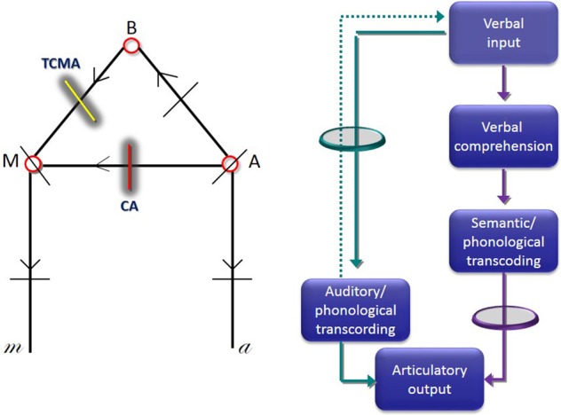
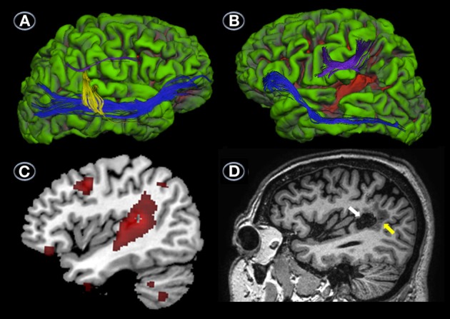
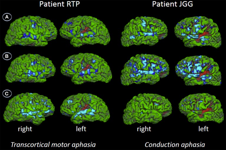
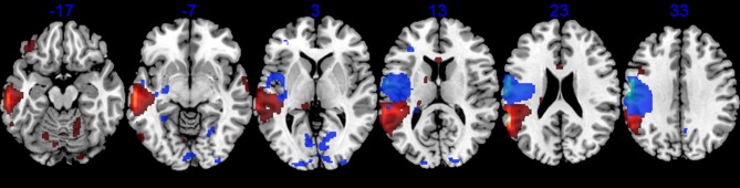
Assessment of brain-damaged subjects presenting with dissociated repetition deficits after selective injury to either the left dorsal or ventral auditory pathways can provide further insight on their respective roles in verbal repetition. We evaluated repetition performance and its neural correlates using multimodal imaging (anatomical MRI, DTI, fMRI, and(18)FDG-PET) in a female patient with transcortical motor aphasia (TCMA) and in a male patient with conduction aphasia (CA) who had small contiguous but non-overlapping left perisylvian infarctions. Repetition in the TCMA patient was fully preserved except for a mild impairment in nonwords and digits, whereas the CA patient had impaired repetition of nonwords, digits and word triplet lists. Sentence repetition was impaired, but he repeated novel sentences significantly better than clichés. The TCMA patient had tissue damage and reduced metabolism in the left sensorimotor cortex and insula. DTI showed damage to the left temporo-frontal and parieto-frontal segments of the arcuate fasciculus (AF) and part of the left ventral stream together with well-developed right dorsal and ventral streams, as has been reported in more than one-third of females. The CA patient had tissue damage and reduced metabolic activity in the left temporoparietal cortex with additional metabolic decrements in the left frontal lobe. DTI showed damage to the left temporo-parietal and temporo-frontal segments of the AF, but the ventral stream was spared. The direct segment of the AF in the right hemisphere was also absent with only vestigial remains of the other dorsal subcomponents present, as is often found in males. fMRI during word and nonword repetition revealed bilateral perisylvian activation in the TCMA patient suggesting recruitment of spared segments of the left dorsal stream and right dorsal stream with propagation of signals to temporal lobe structures suggesting a compensatory reallocation of resources via the ventral streams. The CA patient showed a greater activation of these cortical areas than the TCMA patient, but these changes did not result in normal performance. Repetition of word triplet lists activated bilateral perisylvian cortices in both patients, but activation in the CA patient with very poor performance was restricted to small frontal and posterior temporal foci bilaterally. These findings suggest that dissociated repetition deficits in our cases are probably reliant on flexible interactions between left dorsal stream (spared segments, short tracts remains) and left ventral stream and on gender-dimorphic architecture of the right dorsal stream.
Keywords: conduction aphasia; diffusion tensor tractography; dual dorsal-ventral pathways; functional magnetic resonance imaging; positron emission tomography; repetition; transcortical motor aphasia.
Figures


Similar articles
-
Aphasia.2024 Oct 29. In: StatPearls [Internet]. Treasure Island (FL): StatPearls Publishing; 2025 Jan–. 2024 Oct 29. In: StatPearls [Internet]. Treasure Island (FL): StatPearls Publishing; 2025 Jan–. PMID: 32644741 Free Books & Documents.
-
Repeating with the right hemisphere: reduced interactions between phonological and lexical-semantic systems in crossed aphasia?Front Hum Neurosci. 2013 Oct 18;7:675. doi: 10.3389/fnhum.2013.00675. eCollection 2013. Front Hum Neurosci. 2013. PMID: 24151460 Free PMC article.
-
Damage to ventral and dorsal language pathways in acute aphasia.Brain. 2013 Feb;136(Pt 2):619-29. doi: 10.1093/brain/aws354. Epub 2013 Jan 31. Brain. 2013. PMID: 23378217 Free PMC article.
-
Arcuate fasciculus variability and repetition: the left sometimes can be right.Cortex. 2012 Feb;48(2):133-43. doi: 10.1016/j.cortex.2011.06.014. Epub 2011 Jun 29. Cortex. 2012. PMID: 21802076 Review.
-
Language Learning Variability within the Dorsal and Ventral Streams as a Cue for Compensatory Mechanisms in Aphasia Recovery.Front Hum Neurosci. 2017 Sep 27;11:476. doi: 10.3389/fnhum.2017.00476. eCollection 2017. Front Hum Neurosci. 2017. PMID: 29021751 Free PMC article. Review.
Cited by
-
Transcranial Direct Current Stimulation in Poststroke Aphasia Recovery.Stroke. 2017 Mar;48(3):820-826. doi: 10.1161/STROKEAHA.116.015626. Epub 2017 Feb 7. Stroke. 2017. PMID: 28174328 Free PMC article. Review. No abstract available.
-
Revealing the dual streams of speech processing.Proc Natl Acad Sci U S A. 2016 Dec 27;113(52):15108-15113. doi: 10.1073/pnas.1614038114. Epub 2016 Dec 12. Proc Natl Acad Sci U S A. 2016. PMID: 27956600 Free PMC article.
-
Dissecting the function of networks underpinning language repetition.Front Hum Neurosci. 2014 Oct 2;8:727. doi: 10.3389/fnhum.2014.00727. eCollection 2014. Front Hum Neurosci. 2014. PMID: 25324751 Free PMC article. No abstract available.
-
Developmental conduction aphasia after neonatal stroke.Ann Neurol. 2018 Apr;83(4):664-675. doi: 10.1002/ana.25218. Ann Neurol. 2018. PMID: 29572915 Free PMC article.
References
-
- Albert M. L., Goodglass H., Helm N. A., Rubens A. B., Alexander M. P. (1981). Clinical aspects of dysphasia, in Disorders of Human Communication 2, eds Arnold G. E., Winckel F., Wyke B. D. (Wien; New York: Springer-Verlag; ).
LinkOut - more resources
Full Text Sources
Other Literature Sources