Review
. 2013 Dec 20:4:380.
doi: 10.3389/fphys.2013.00380.

Acid-base transport in pancreas-new challenges

Affiliations
Review

Acid-base transport in pancreas-new challenges

Ivana Novak et al. Front Physiol. .

Abstract

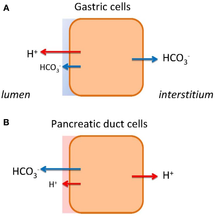
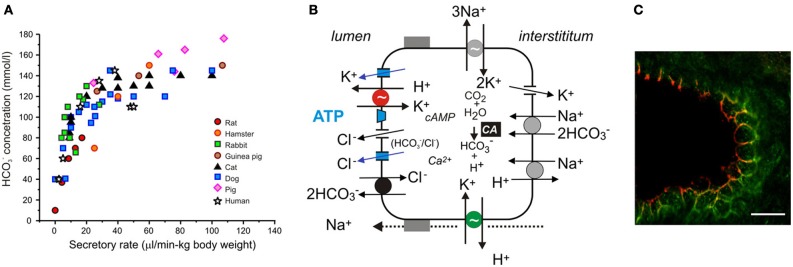
Along the gastrointestinal tract a number of epithelia contribute with acid or basic secretions in order to aid digestive processes. The stomach and pancreas are the most extreme examples of acid (H(+)) and base (HCO(-) 3) transporters, respectively. Nevertheless, they share the same challenges of transporting acid and bases across epithelia and effectively regulating their intracellular pH. In this review, we will make use of comparative physiology to enlighten the cellular mechanisms of pancreatic HCO(-) 3 and fluid secretion, which is still challenging physiologists. Some of the novel transporters to consider in pancreas are the proton pumps (H(+)-K(+)-ATPases), as well as the calcium-activated K(+) and Cl(-) channels, such as KCa3.1 and TMEM16A/ANO1. Local regulators, such as purinergic signaling, fine-tune, and coordinate pancreatic secretion. Lastly, we speculate whether dys-regulation of acid-base transport contributes to pancreatic diseases including cystic fibrosis, pancreatitis, and cancer.

Keywords: ANO1; H+-K+-ATPase; IK; KCa3.1; TMEM16A; bicarbonate transport; pancreatic duct; proton transport.


Figures

Figure 1
Figure 1
HCO3 and H+ transport in gastric cells (A) and pancreatic duct cells (B). The models show schematically different types of epithelia as single cells. The transport of H+ or HCO3 to the bulk luminal fluid is shown with large arrows. The small arrows on luminal side indicate HCO3 and H+ secretions to the mucosal buffer zone. Flux of HCO3 and H+ to the interstititum/blood side indicates expected alkaline or acid tides.
Figure 2
Figure 2
Acid/base transport in pancreas. (A) The relation between secretory rates and HCO3 concentrations in pancreatic juice of various species. Secretions were stimulated by secretin and secretory rates were corrected for body weights. (B) The model of ion transport in a secreting pancreatic duct cell with novel transporters, channels and luminal purinergic signaling and receptors indicated in color and discussed in the review. Intracellular HCO3 is derived from CO2 through the action of carbonic anhydrase (CA) and from HCO3 uptake via the electrogenic Na+–HCO3 cotransporter (pNBC, NBCe1). H+ is extruded at basolateral membrane by the Na+/H+ exchanger (NHE1). HCO3 efflux across the luminal membrane is mediated by the electrogenic Cl/HCO3 exchanger (SLC26A6), and under certain conditions, through Cl channels. The luminal Cl channels are CFTR and TMEM16A (see text). There are a number of K+ channels expressed on the luminal and basolateral membranes, e.g., KCa3.1, KCa1.1, KCNQ1 (see text). The luminal and basolateral H+-K+-ATPases are indicated in red and green, and supposedly contribute to the luminal buffer zone and the H+ efflux to intersititum, respectively. Other ion channels and transporters, such as NHE3, SLC26A3, NBC3, NKCC1, and aquaporins have a differential distribution in the duct tree and for simplicity are not included in the model. (C) Immunolocalization of the gastric (red) and non-gastric (green) H+-K+ pumps in rat pancreatic duct. The bar is 20 μm. Modified from Novak et al., .

References

    1. Almassy J., Won J. H., Begenisich T. B., Yule D. I. (2012). Apical Ca2+-activated potassium channels in mouse parotid acinar cells. J. Gen. Physiol. 139, 121–133 10.1085/jgp.201110718 - DOI - PMC - PubMed
    1. Alvarez C., Regan J. P., Merianos D., Bass B. L. (2004). Protease-activated receptor-2 regulates bicarbonate secretion by pancreatic duct cells in vitro. Surgery 136, 669–676 10.1016/j.surg.2004.01.018 - DOI - PubMed
    1. Ashizawa N., Endoh H., Hidaka K., Watanabe M., Fukumoto S. (1997). Three-dimensional structure of the rat pancreatic duct in normal and inflammated pancreas. Microsc. Res. Tech. 37, 543–556 10.1002/(SICI)1097-0029(19970601)37:5/6<543::AID-JEMT15>3.3.CO;2-Y - DOI - PubMed
    1. Ashley S. W., Schwarz M., Alvarez C., Nguyen T. N., Vdovenko A., Reber H. A. (1994). Pancreatic interstitial pH regulation: effects of secretory stimulation. Surgery 115, 503–509 - PubMed
    1. Behrendorff N., Floetenmeyer M., Schwiening C., Thorn P. (2010). Protons released during pancreatic acinar cell secretion acidify the lumen and contribute to pancreatitis in mice. Gastroenterology 139, 1711-20, 1720.e1-5. 10.1053/j.gastro.2010.07.051 - DOI - PubMed

LinkOut - more resources