

Real-time magnetic resonance imaging (MRI) during active wrist motion--initial observations
- PMID: 24391865
- PMCID: PMC3877133
- DOI: 10.1371/journal.pone.0084004
Real-time magnetic resonance imaging (MRI) during active wrist motion--initial observations
Abstract
Background: Non-invasive imaging techniques such as magnetic resonance imaging (MRI) provide the ability to evaluate the complex anatomy of bone and soft tissues of the wrist without the use of ionizing radiation. Dynamic instability of wrist--occurring during joint motion--is a complex condition that has assumed increased importance in musculoskeletal medicine. The objective of this study was to develop an MRI protocol for evaluating the wrist during continuous active motion, to show that dynamic imaging of the wrist is realizable, and to demonstrate that the resulting anatomical images enable the measurement of metrics commonly evaluated for dynamic wrist instability.
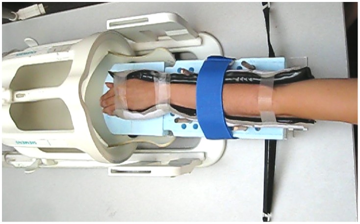
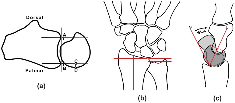
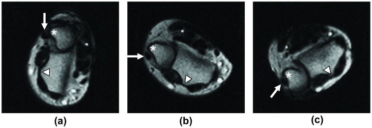
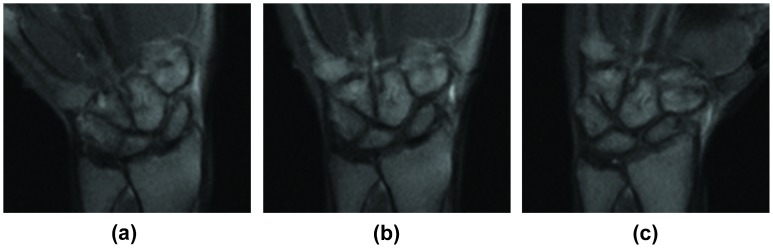
Methods: A 3-Tesla "active-MRI" protocol was developed using a bSSFP sequence with 475 ms temporal resolution for continuous imaging of the moving wrist. Fifteen wrists of 10 asymptomatic volunteers were scanned during active supination/pronation, radial/ulnar deviation, "clenched-fist", and volarflexion/dorsiflexion maneuvers. Two physicians evaluated distal radioulnar joint (DRUJ) congruity, extensor carpi ulnaris (ECU) tendon translation, the scapholunate (SL) interval, and the SL, radiolunate (RL) and capitolunate (CL) angles from the resulting images.
Results: The mean DRUJ subluxation ratio was 0.04 in supination, 0.10 in neutral, and 0.14 in pronation. The ECU tendon was subluxated or translated out of its groove in 3 wrists in pronation, 9 wrists in neutral, and 11 wrists in supination. The mean SL interval was 1.43 mm for neutral, ulnar deviation, radial deviation positions, and increased to 1.64 mm during the clenched-fist maneuver. Measurement of SL, RL and CL angles in neutral and dorsiflexion was also accomplished.
Conclusion: This study demonstrates the initial performance of active-MRI, which may be useful in the investigation of dynamic wrist instability in vivo.
Conflict of interest statement
Figures
References
-
- Garcia-Elias M (1999) Position Statement: Definition of Carpal Instability: The Anatomy and Biomechanics Committee of the International Federation of Societies for Surgery of the Hand2. The Journal of Hand Surgery 24: 866–867. - PubMed
-
- Garcia-Elias M (1997) The treatment of wrist instability. The Journal of Bbone and Joint Surgery British volume 79: 684. - PubMed
-
- Taleisnik J (1988) Current concepts review. Carpal instability. J Bone Joint Surg Am 70: 1262–1268. - PubMed
-
- Watson HK, Ryu J, Akelman E (1986) Limited triscaphoid intercarpal arthrodesis for rotatory subluxation of the scaphoid. J Bone Joint Surg A 68: 345–349. - PubMed
-
- Theumann NH, Etechami G, Duvoisin B, Wintermark M, Schnyder P, et al. (2006) Association between Extrinsic and Intrinsic Carpal Ligament Injuries at MR Arthrography and Carpal Instability at Radiography: Initial Observations. Radiology 238: 950–957. - PubMed
Publication types
MeSH terms
Grants and funding
LinkOut - more resources
Full Text Sources
Other Literature Sources
Medical
