. 2013 Dec 31;8(12):e84256.
doi: 10.1371/journal.pone.0084256. eCollection 2013.

Exosomes derived from mesenchymal stem cells suppress angiogenesis by down-regulating VEGF expression in breast cancer cells

Affiliations

Exosomes derived from mesenchymal stem cells suppress angiogenesis by down-regulating VEGF expression in breast cancer cells

Jong-Kuen Lee et al. PLoS One. .

Abstract

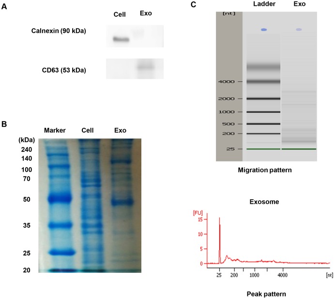
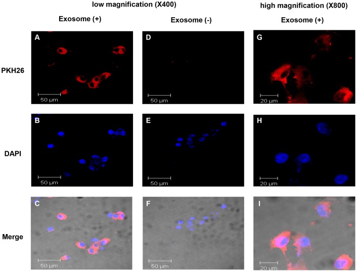
Exosomes are small membrane vesicles released by a variety of cell types. Exosomes contain genetic materials, such as mRNAs and microRNAs (miRNAs), implying that they may play a pivotal role in cell-to-cell communication. Mesenchymal stem cells (MSCs), which potentially differentiate into multiple cell types, can migrate to the tumor sites and have been reported to exert complex effects on tumor progression. To elucidate the role of MSCs within the tumor microenvironment, previous studies have suggested various mechanisms such as immune modulation and secreted factors of MSCs. However, the paracrine effects of MSC-derived exosomes on the tumor microenvironment remain to be explored. The hypothesis of this study was that MSC-derived exosomes might reprogram tumor behavior by transferring their molecular contents. To test this hypothesis, exosomes from MSCs were isolated and characterized. MSC-derived exosomes exhibited different protein and RNA profiles compared with their donor cells and these vesicles could be internalized by breast cancer cells. The results demonstrated that MSC-derived exosomes significantly down-regulated the expression of vascular endothelial growth factor (VEGF) in tumor cells, which lead to inhibition of angiogenesis in vitro and in vivo. Additionally, miR-16, a miRNA known to target VEGF, was enriched in MSC-derived exosomes and it was partially responsible for the anti-angiogenic effect of MSC-derived exosomes. The collective results suggest that MSC-derived exosomes may serve as a significant mediator of cell-to-cell communication within the tumor microenvironment and suppress angiogenesis by transferring anti-angiogenic molecules.


Conflict of interest statement

Competing Interests: The authors have declared that no competing interests exist.

Figures

Figure 1
Figure 1. Characterization of MSC-derived exosomes.
(A) Western blotting was performed with MSCs (Cell) or MSC-derived exosomes (Exo). Calnexin expression in MSCs and CD63 expression in MSC-derived exosomes were detected. (B) Protein was isolated from MSCs and MSC-derived exosomes. An equivalent amount (50 ug) of protein from MSCs and MSC-derived exosomes was loaded and run on a 10% SDS gel and stained with Coomassie blue. (C) RNA was extracted from MSC-derived exosomes and analyzed by a Bioanalyzer. Representative bioanalyzer profile of the RNA contained in MSC-derived exosomes showed that the ribosomal subunits 28S and 18S were barely detectable.
Figure 2
Figure 2. Cellular internalization of MSC-derived exosomes into 4T1 cells.
4T1 cells were incubated with 25 μg of MSC-derived exosomes that were labeled with PKH26 (red) for 24 h. 4T1 cells were also incubated with PKH26 without exosomes as a negative control to observe carryover of PKH26. Low magnification images of 4T1 cells incubated with exosomes (A–C), or negative controls without exosomes (D–F) are shown (×400). High magnification images of 4T1 cells incubated with exosomes (G–I) are shown (×800).
Figure 3
Figure 3. Down-regulation of VEGF expression in 4T1 cells by MSC-derived exosomes.
(A) 4T1 cells were incubated with various concentrations of MSC-derived exosomes (25 μg/ml, 50 μg/ml, and 100 μg/ml) or carrier control (PBS) for 48 h. The mRNA levels of VEGF were evaluated using qRT-PCR. (B) The levels of secreted VEGF in the conditioned media from 4T1 cells that were treated with various concentrations of MSC-derived exosomes (25 μg/ml, 50 μg/ml, and 100 μg/ml) or carrier control (PBS) for 24 h were estimated by enzyme-linked immunosorbent assay. (C) The mRNA levels of VEGFR-1 in 4T1 cells that were stimulated with MSC-derived exosomes were analyzed by qRT-PCR. The values are presented as the mean ± D; n = 3 for each group. Significant differences were evaluated using an unpaired two-tailed Student's t-test. NS; Not significant, *P<0.05, **P<0.01, ***P<0.001 compared with control.
Figure 4
Figure 4. Transfer of miR-16 via MSC-derived exosomes.
(A) qRT-PCR was used to measure the levels of miR-16 in MSCs and MSC-derived exosomes. (B) 4T1 cells were incubated with MSC-derived exosomes and α-amanitin (experimental sample) or α-amanitin alone (negative control). Transfer of miR-16 was determined by qRT-PCR and positive values indicate transfer of miR-16. (C) 4T1 cells were incubated with various concentrations of MSC-derived exosomes (25 μg/ml, 50 μg/ml, and 100 μg/ml) or carrier control (PBS) for 48 h and miR-16 levels were evaluated. (D) 4T1 cells were transfected with miR-16 inhibitor and incubated with MSC-derived exosomes (100 μg/ml) or carrier control (PBS) for 24 h. qRT-PCR was conducted to evaluate the VEGF mRNA expression. The values are presented as the mean ± SD; n = 3 for each group. Significant differences were evaluated using an unpaired two-tailed Student's t-test. NS; Not significant, *P<0.05, **P<0.01, ***P<0.001 compared with control.
Figure 5
Figure 5. MSC-derived exosomes inhibit proliferation and migration of SVEC cells in vitro.
(A) 2.0×10³ SVEC cells were incubated with the conditioned media from 4T1 cells that were treated with various concentrations of MSC-derived exosomes or carrier control (PBS). Cell proliferation rates were determined by an EZ-Cytox cell viability assay kit. (B) SVEC cells transwell migration assay was performed in the presence of the conditioned media from 4T1 cells that were treated with various concentrations of MSC-derived exosomes or carrier control (PBS) in the lower chambers. Serum-starved SVEC cells were added to the upper chamber and incubated for 24 h to allow cell migration through the membrane. The membranes were stained with crystal violet and cell migration was analyzed by Image J. (C) SVEC cells were scratched and incubated with the conditioned media from 4T1 cells stimulated with MSC-derived exosomes (100 μg/ml) or vehicle control (PBS) for 24 h. In order to neutralize VEGF derived from 4T1 cells, anti-VEGF antibodies (20 μg/ml) were added to the conditioned media. Photographs were taken immediately and 24 h after wounding (data not shown) and analyzed by Studio Lite, version 1.0. (D) SVEC cells were serum-starved for 24 h and 2×10⁴ SVEC cells were seeded in a Matrigel-coated well. The cells were treated with conditioned media collected from 4T1 cells stimulated with MSC-derived exosomes (100 μg/ml) or carrier control (PBS) for 24 h and viewed under a microscope. The values are presented as the mean ± SD; n = 3 for each group. Significant differences were evaluated using an unpaired two-tailed Student's t-test. *P<0.05, **P<0.01, ***P<0.001 compared with control.
Figure 6
Figure 6. MSC-derived exosomes suppress angiogenesis in vivo.
(A) BALB/c mice received subcutaneous injections of 100 μl PBS per mouse containing 2×10⁵ 4T1 cells alone or 2×10⁵ 4T1 cells mixed with 100 μg of MSC-derived exosomes or 2×10⁵ 4T1 cells mixed with 200 μg of MSC-derived exosomes. The tumor sizes of mice in the groups were measured with a caliper three times a week from 20 days after tumor challenges. (B) The tumor weight was measured. (C) VEGF mRNA expressions in tumor tissues were analyzed by using qRT-PCR. (D) Immunohistochemical features of the tumor tissues were shown. Formalin-fixed paraffin sections were stained with anti-VEGF and anti-CD31 antibodies. (E) The mean number of vessels in tumor histologic sections was quantified. The values are presented as the mean ± SD; n = 4 for each group. Significant differences were evaluated using an unpaired two-tailed Student's t-test. *P<0.05, **P<0.01, ***P<0.001 compared with control.

References

    1. Raposo G, Nijman HW, Stoorvogel W, Liejendekker R, Harding CV, et al. (1996) B lymphocytes secrete antigen-presenting vesicles. J Exp Med 183: 1161–1172. - PMC - PubMed
    1. Viaud S, Ullrich E, Zitvogel L, Chaput N (2008) Exosomes for the treatment of human malignancies. Horm Metab Res 40: 82–88. - PubMed
    1. Valadi H, Ekstrom K, Bossios A, Sjostrand M, Lee JJ, et al. (2007) Exosome-mediated transfer of mRNAs and microRNAs is a novel mechanism of genetic exchange between cells. Nat Cell Biol 9: 654–659. - PubMed
    1. Camussi G, Deregibus MC, Bruno S, Grange C, Fonsato V, et al. (2011) Exosome/microvesicle-mediated epigenetic reprogramming of cells. Am J Cancer Res 1: 98–110. - PMC - PubMed
    1. Winter J, Jung S, Keller S, Gregory RI, Diederichs S (2009) Many roads to maturity: microRNA biogenesis pathways and their regulation. Nat Cell Biol 11: 228–234. - PubMed

Publication types

MeSH terms