Comparative Study
. 2013 Dec 31;8(12):e84272.
doi: 10.1371/journal.pone.0084272. eCollection 2013.

Comparative characteristics of porous bioceramics for an osteogenic response in vitro and in vivo

Affiliations
Comparative Study

Comparative characteristics of porous bioceramics for an osteogenic response in vitro and in vivo

Hye-Rim Lee et al. PLoS One. .

Abstract

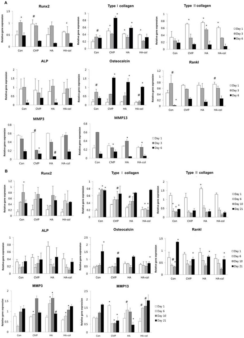
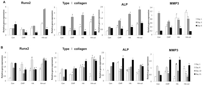
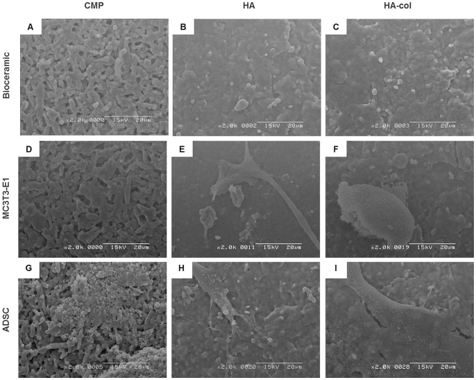
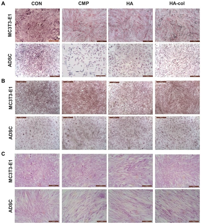
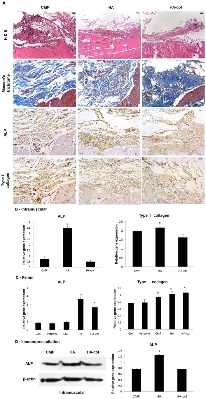
Porous calcium phosphate ceramics are used in orthopedic and craniofacial applications to treat bone loss, or in dental applications to replace missing teeth. The implantation of these materials, however, does not induce stem cell differentiation, so suitable additional materials such as porous calcium phosphate discs are needed to influence physicochemical responses or structural changes. Rabbit adipose-derived stem cells (ADSC) and mouse osteoblastic cells (MC3T3-E1) were evaluated in vitro by the MTT assay, semi-quantitative RT-PCR, and immunoblotting using cells cultured in medium supplemented with extracts from bioceramics, including calcium metaphosphate (CMP), hydroxyapatite (HA) and collagen-grafted HA (HA-col). In vivo evaluation of the bone forming capacity of these bioceramics in rat models using femur defects and intramuscular implants for 12 weeks was performed. Histological analysis showed that newly formed stromal-rich tissues were observed in all the implanted regions and that the implants showed positive immunoreaction against type I collagen and alkaline phosphatase (ALP). The intramuscular implant region, in particular, showed strong positive immunoreactivity for both type I collagen and ALP, which was further confirmed by mRNA expression and immunoblotting results, indicating that each bioceramic material enhanced osteogenesis stimulation. These results support our hypothesis that smart bioceramics can induce osteoconduction and osteoinduction in vivo, although mature bone formation, including lacunae, osteocytes, and mineralization, was not prominent until 12 weeks after implantation.


Conflict of interest statement

Competing Interests: The authors have declared that no competing interests exist.

Figures

Figure 1
Figure 1. Effect of bioceramics on the proliferation of MC3T3-E1 cells (A) and ADSC (B).
Bioceramics (CMP, HA and HA-col) were incubated for 24 h in standard medium (DMEM and α-MEM with 10% FBS and antibiotics) at 37°C, 5% CO2 in 6-well dishes. 2×10² cells (ADSC and MC3T3-E1) were seeded in 96-well dishes, and the wells were filled with 100 μl medium. *p<0.01 versus the control.
Figure 2
Figure 2. Quantitative analysis of the expression of genes involved in bone formation and bone resorption.
mRNA expression of Runx2, type I collagen, type II collagen, ALP, osteocalcin, Rankl, MMP3, and MMP13 genes in MC3T3-E1 cells at 1, 3, and 6 d (A), and ADSC at 1, 6, 10, and 21 d (B) of culture in bioceramics release medium. Compared to the control, the expression level of type I collagen and osteocalcin of MC3T3-E1 cells was increased and those of Runx2, Rankl, MMP3 and MMP13 gene expression were decreased, each of which occurred in a time-dependent manner. Osteoblast dependent increases in osteogenic gene expression of Runx2, type I collagen, ALP and osteocalcin were observed for each bioceramic in ADSC cultures. Data are presented as mean ± standard deviation (n = 3). #p<0.05, *p<0.01.
Figure 3
Figure 3. Immunoblotting for protein expression of osteogenic molecules in MC3T3-E1 cells (A) and ADSC (B).
Relative ratios of Runx2, Type I collagen, ALP, MMP3 to β-actin were measured with Image J software. Data are representative of at least two experiments. Values are the mean ± standard deviation. Statistical difference as compared to corresponding control group. #p<0.05, *p<0.01.
Figure 4
Figure 4. Scanning electron microscope observation of cell adhesion on bioceramic surfaces at 6 d of MC3T3-E1 cell culture, and at 10 d of ADSC culture.
(A–C) Bioceramics only; (D–F) ADSC culture on bioceramic; (G–I) MC3T3-E1 culture on bioceramic. At 6 d of MC3T3-E1 culture, the cells were attached, exhibiting polygonal shapes on HA (E) and HA-col (F). At 10 d after ADSC seeding, the SEM images showed that cells were well attached on the surfaces of HA (H) and HA-col (I) with filopodia. Bioceramic: CMP, HA and HA-col. Scale Bar: 20 mm.
Figure 5
Figure 5. Cytochemical analysis shows extracellular matrix collagen and mineralization in ADSC cultures at 10 d, and MC3T3-E1 cell cultures at 6 d in bioceramic medium.
Cells were plated in 6-well plates at a seeding density of 1×10⁶ cells/well, and were cultured for 6 and 10 d in a media containing ceramics. (A) Picrosirius staining shows the presence of type I collagen (red, orange, and yellow) in cell layers. (B) Alizarin Red S and (C) Von Kossa staining shows mineralization of the extracellular matrix.
Figure 6
Figure 6. Results of radiopacity for the newly formed tissues after bioceramic implantation. Representative radiographs (A) and histological analysis (B) of a femur with bioceramics implantation for 12 weeks.
(A) Density of the bone was analyzed by X-ray, and radiopacity levels were measured at the same area (red rectangle) from the bioceramics implanted region to the opposite side. The graph shows the relative opacity of X-ray films, which indicates the kind of implanted bioceramics. The stronger the radiopaque image in the red dashed rectangle, the higher the bone density. It is clear that the CMP sample exhibits higher radiopacity than the others. Vetbond, HA, and HA-col groups were at the same relative opacity, which was higher than that in the control. *p<0.01 versus the control. (B) The cortical defect on the femur of all groups showed newly formed eosinophilic connective tissues. At higher magnification, the composition and density of the newly formed tissues of bioceramics were different from the control and Vetbond samples. Bioceramic-implanted groups revealed marked compact structures when compared to the others, which corresponds with the immunoreactivity of ALP and type I collagen. Representative H&E stained sections of bioceramic implants from 12 weeks after surgery. H&E, Magnification, ×100 and ×200. Immunohistochemistry for ALP and type I collagen, Mayer's hematoxylin counter staining, Magnification, ×200.
Figure 7
Figure 7. Histological analysis at 12 weeks post-implantation intramuscularly (A).
In H&E staining, peripheral regions of the bioceramic-implanted muscles showed variable degrees of newly formed eosinophilic connective tissue deposition, which show a positive reaction to trichrome staining indicating a collagen-rich stroma. All bioceramic-implanted groups showed positive immunohistochemistry reaction to ALP and type I collagen but differences of positive intensity between each group were not obvious. Immunohistochemistry, Mayer's hematoxylin counter staining, Magnification, ×200. RT-PCR (B, C) for gene and immunoblotting for protein (D) expression of osteogenic molecules in the intramuscularis and cortical defects. (B, C) Formalin fixed paraffin-embedded (FFPE) muscular and femur tissue RNA was extracted by All prep DNA/RNA FFPE kit. In muscles, mRNA expression of ALP was increased to a greater degree in the HA group than in others, with similar results observed by immunoblotting as shown in Figure 6B. Type I collagen was up-regulated in the CMP and HA groups. (D) Expression of ALP was higher in the HA group than in the others, however, there was no significant difference in the expression level of type I collagen among all groups. Protein extraction from FFPE muscular tissues by Q proteome FFPE tissue kit for immunoblotting. Values are the mean ± standard deviation. #p<0.05, *p<0.01 versus the CMP group.

References

    1. Wang C, Duan Y, Markovic B, Barbara J, Howlett CR, et al. (2004) Phenotypic expression of bone-related genes in osteoblasts grown on calcium phosphate ceramics with different phase compositions. Biomaterials 25: 2507–2514 10.1016/j.biomaterials.2003.09.035.PubMed:14751735 - DOI - PubMed
    1. Akita S, Tamai N, Myoui A, Nishikawa M, Kaito T, et al. (2004) Capillary vessel network integration by inserting a vascular pedicle enhances bone formation in tissue-engineered bone using interconnected porous hydroxyapatite ceramics. Tissue Eng 10: 789–795 10.1089/1076327041348338.PubMed:15265296 - DOI - PubMed
    1. Ripamonti U (1991) The morphogenesis of bone in replicas of porous hydroxyapatite obtained from conversion of calcium carbonate exoskeletons of coral. J Bone Joint Surg Am 73: 692–703. PubMed: 1675218. - PubMed
    1. Ogata K, Imazato S, Ehara A, Ebisu S, Kinomoto Y, et al. (2005) Comparison of osteoblast responses to hydroxyapatite and hydroxyapatite/soluble calcium phosphate composites. J Biomed Mater Res A 72: 127–135 10.1002/jbm.a.30146.PubMed:15625683 - DOI - PubMed
    1. Kitsugi T, Yamamuro T, Nakamura T, Oka M (1995) Transmission electron microscopy observations at the interface of bone and four types of calcium phosphate ceramics with different calcium/phosphorus molar ratios. Biomaterials 16: 1101–1107 10.1016/0142-9612(95)98907-V.PubMed:8519932 - DOI - PubMed

Publication types

MeSH terms

LinkOut - more resources