. 2013 Dec 31;8(12):e85737.
doi: 10.1371/journal.pone.0085737. eCollection 2013.

Syndecan-1 (CD138) modulates triple-negative breast cancer stem cell properties via regulation of LRP-6 and IL-6-mediated STAT3 signaling

Affiliations

Syndecan-1 (CD138) modulates triple-negative breast cancer stem cell properties via regulation of LRP-6 and IL-6-mediated STAT3 signaling

Sherif A Ibrahim et al. PLoS One. .

Abstract

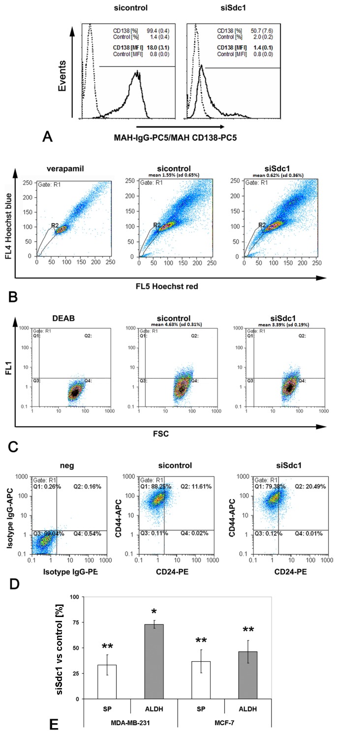
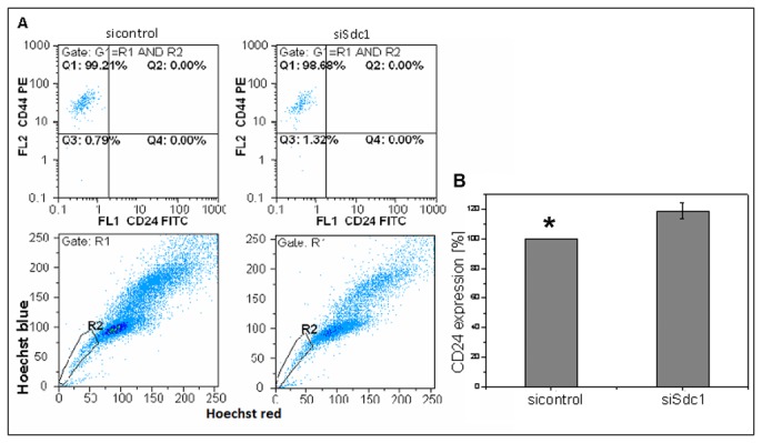
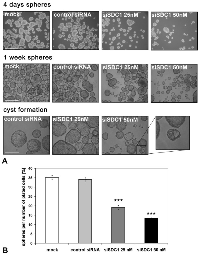
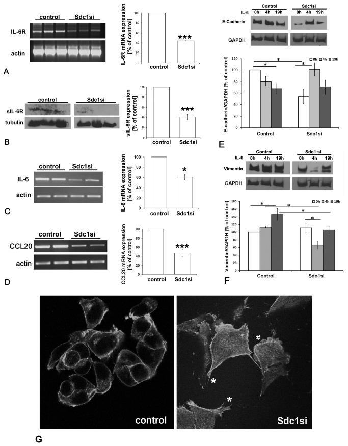
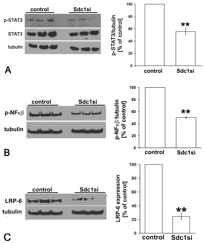
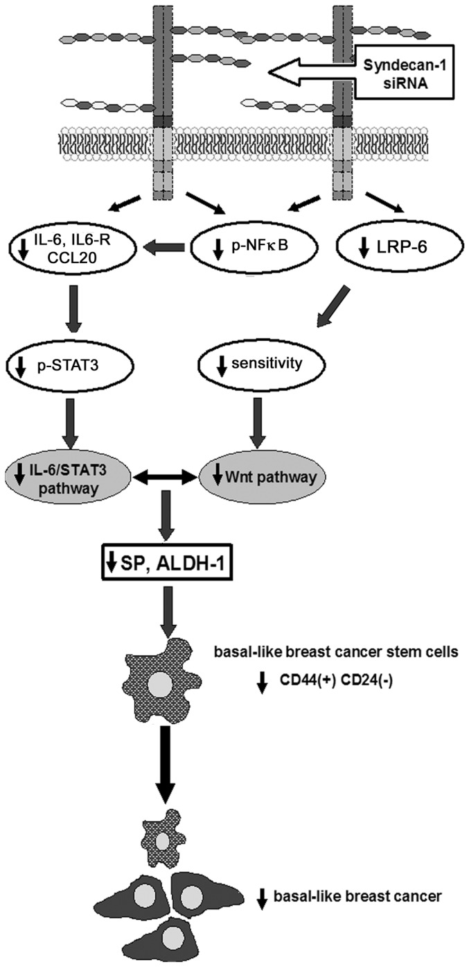
Syndecan-1 (CD138), a heparan sulfate proteoglycan, acts as a coreceptor for growth factors and chemokines and is a molecular marker associated with epithelial-mesenchymal transition during development and carcinogenesis. Resistance of Syndecan-1-deficient mice to experimentally-induced tumorigenesis has been linked to altered Wnt-responsive precursor cell pools, suggesting a potential role of Syndecan-1 in breast cancer cell stem function. However, the precise molecular mechanism is still elusive. Here, we decipher the functional impact of Syndecan-1 knockdown using RNA interference on the breast cancer stem cell phenotype of human triple-negative MDA-MB-231 and hormone receptor-positive MCF-7 cells in vitro employing an analytical flow cytometric approach. Successful Syndecan-1 siRNA knockdown was confirmed by flow cytometry. Side population measurement by Hoechst dye exclusion and Aldehyde dehydrogenase-1 activity revealed that Syndecan-1 knockdown in MDA-MB-231 cells significantly reduced putative cancer stem cell pools by 60% and 27%, respectively, compared to controls. In MCF-7 cells, Syndecan-1 depletion reduced the side population by 40% and Aldehyde dehydrogenase-1 by 50%, repectively. In MDA-MB-231 cells, the CD44(+)CD24(-/low) phenotype decreased significantly by 6% upon siRNA-mediated Syndecan-1 depletion. Intriguingly, IL-6, its receptor sIL-6R, and the chemokine CCL20, implicated in regulating stemness-associated pathways, were downregulated by >40% in Syndecan-1-silenced MDA-MB-231 cells, which showed a dysregulated response to IL-6-induced shifts in E-cadherin and vimentin expression. Furthermore, activation of STAT-3 and NFkB transcription factors and expression of a coreceptor for Wnt signaling, LRP-6, were reduced by >45% in Syndecan-1-depleted cells compared to controls. At the functional level, Syndecan-1 siRNA reduced the formation of spheres and cysts in MCF-7 cells grown in suspension culture. Our study demonstrates the viability of flow cytometric approaches in analyzing cancer stem cell function. As Syndecan-1 modulates the cancer stem cell phenotype via regulation of the Wnt and IL-6/STAT3 signaling pathways, it emerges as a promising novel target for therapeutic approaches.


Conflict of interest statement

Competing Interests: The authors have declared that no competing interests exist.

Figures

Figure 1
Figure 1. Flow cytometric analysis of putative cancer stem cell pools in control and Syndecan-1-silenced MDA-MB-231 breast cancer cells.
Syndecan-1 silencing reduces the SP phenotype, ALDH1 activity, and the CD44(+)/CD24(-/low) phenotype. A) Confirmation of Syndecan-1 siRNA knockdown by flow cytometry. The percentage of marker-positive cells and the mean fluorescent intensity of events (mean (SD), n=4) are depicted on the plots. (Dotted lines: mouse-IgG1-PC5 control; solid lines: anti-CD138-PC5; marker for percentage of CD138-positive cells; MFI = median fluorescent intensity). B) Side population analysis. Control and Syndecan-1-depleted cells were incubated with the Hoechst 33342 dye in the presence or absence of 50 µM verapamil, and subjected to flow cytometry. Gate R1 shows SP cells. C) Example of the identification of putative cancer stem cells displaying aldehyde dehydrogenase (ALDH) activity. Control and Syndecan-1 knockdown cells were incubated with fluorescent ALDH substrate in the presence or absence of the inhibitor diethylaminobenzaldehyde (DEAB), followed by flow cytometric analysis. D) Example of flow cytometric analysis for the cell surface expression of CD44 and CD24 protein. Control and Syndecan-1 knockdown cells were incubated with isotype IgG-PE and-APC, CD24-PE and CD44-APC antibodies followed by sorting the cells by flow cytometry into left lower quadrant CD44(-)CD24(-),left upper quadrant CD44(+)CD24(-), right lower quadrant CD44(-)CD24(+), right upper quadrant CD44(+)CD24(+). A-D) Representative examples of n≥3 experiments. E) Quantitative analysis of SP and ALDH measurements in Syndecan-1 and control siRNA treated MDA-MB-231 and MCF-7 cells. Syndecan-1 siRNA knockdown results in a significant decrease of the SP and ALDH activity. Data are expressed as mean percentage +/- SEM relative to the controls (set to 100%). (n=3-5, *=P<0.05, **=P<0.01).
Figure 2
Figure 2. Characterization of SP cells concerning their CD44/CD24 expression.
The combination of SP- and CD44/CD24 measurement revealed a proportion of 99.2% of SP cells with the CD44+/CD24(-/low) phenotype after control siRNA treatment while the Syndecan-1 knockdown approach showed that this proportion decreased only slightly to 98.7% (A). Influence of Syndecan-1 knockdown on CD24 expression. Compared to the control-siRNA treated cells, the Syndecan-1-siRNA transfected cells showed a 20% (±8.0%) increase in CD24 expression (*=P<0.05, n=3) (B).
Figure 3
Figure 3. Syndecan-1 silencing impairs the formation of spheres and differentiation into cysts in MCF-7 cells.
A) MCF-7 with sphere formation capacity were enriched for and transfected with a control siRNA or Syndecan-1 siRNA, followed by placing the cells in non-adherent culture conditions that promote sphere formation from single cells. After 4 days in suspension without siRNA it’s evident that Syndecan-1 knockdown is affecting proliferation. At this step it is already visible that the spheres are not only smaller but also more irregular compared to controls (upper panel). After 1 week, the spheres formed by Syndecan-1 transfected MCF-7 are less abundant, smaller and there are much more aggregates compared to controls (central panel). After 1 week in suspension culture, MCF-7 cells start to form cysts (bottom panel). The Syndecan-1-transfected cells show a drastically reduced cyst formation capability. The insert shows a Syndecan-1-depleted sphere that is generating a small “sphere/cyst hybrid”. Scale bar = 500µm (upper, central panels); 200µm (lower panel). B) Quantitative analysis of the sphere formation efficiency in control and Syndecan-1 siRNA-treated MCF-7 cells. Sphere number was determined 1 week after plating 3.000 transfected cells. Syndecan-1 siRNA-treatment results in a significant reduction of sphere formation efficiency (P<0.001, n=6).
Figure 4
Figure 4. siRNA-mediated knockdown of Syndecan-1 downregulates expression of IL-6R, IL-6 and CCL20 and dysregulates epithelial and mesenchymal marker protein expression in MDA-MB-231 breast cancer cells.
A) left panel: RT-PCR analysis of IL-6R expression in MDA-MB-231 cells subjected to Syndecan-1 siRNA knockdown. Following total RNA isolation, mRNA was reverse transcribed and used as a template for PCR amplification of IL-6R. Right panel: PCR band intensities were normalized for actin expression and the data were analyzed using the paired Student's t-test. B) Left panel: Western blot analysis reveals reduction of IL-6R following Syndecan-1 silencing. Lysates of control and Syndecan-1 silenced cells were collected and 30-50µg protein/lane was immunoblotted and probed with sIL-6R antibody. Right panel: Immunoblot band intensities were normalized for tubulin expression and the data were analyzed using the paired Student's t-test. Data shown are triplicates from a single experiment representative of three independent experiments. C) left panel: RT-PCR analysis of IL-6 expression. Right panel: semiquantitative densitometric analysis (see panel A). D) left panel: RT-PCR analysis of CCL20 expression. Right panel: semiquantitative densitometric analysis (see panel A). *=p<0.05, ***=p<0.001, n≥3, error bars=SEM. E,F) The influence of IL-6 treatment on the expression of the epithelial marker E-cadherin (E) and the mesenchymal marker vimentin (F) was studied by Western blotting. Cells were stimulated by 50ng/ml IL-6 24h after transfection with siRNA for 4h and 19h. In control cells, IL-6 treatment for 19h promoted EMT. Syndecan-1 depletion resulted in significant downregulation of E-cadherin expression. IL-6 treatment of Syndecan-1 depleted cells for 4h resulted in marker expression changes suggestive of enhanced mesenchymal-to-epithelial transition. (E,F) Upper panels = representative Western blots, lower panels = quantitative analysis. n≥3,*=P<0.05. G) Confocal immunofluorescence microscopy of phalloidin-labeled actin filaments reveals increased formation of actin stress fibers, filopodia (*) and lamellopodia (#) in Syndecan-1 siRNA-treated compared to control siRNA treated MCF-7 cells.
Figure 5
Figure 5. Syndecan−1 modulates activation of the STAT3 and NFκB signaling pathways and expression of LRP-6 in MDA−MB−231 breast cancer cells.
Lysates of control and Syndecan−1 silenced cells were collected and 30-50µg protein/lane was immunoblotted and probed with the indicated antibodies. A) Western blot analysis reveals reduction of the phosphorylated form of STAT3 in Syndecan−1−silenced cells compared to controls. B) Syndecan−1 depletion leads to a significant reduced activation of NFκB. Immunoblot band intensities were normalized for tubulin expression and the data were analyzed using the paired Student's t-test. Data shown are triplicates from a single experiment representative of three independent experiments. **=p≤0.01, n≥3, error bars=SEM. C) Lysates of control and Syndecan−1 silenced cells were collected and 30-50µg protein/lane was immunoblotted and probed with an antibody recognizing LRP-6 (left panel). Immunoblot band intensities were normalized for tubulin expression and the data were analyzed using the paired Student's t-test (right panel). Data shown are triplicates from a single experiment representative of three independent experiments. **=p≤0.01, n≥3, error bars=SEM.
Figure 6
Figure 6. Syndecan-1 modulates triple-negative breast cancer stem cell properties via the IL-6/STAT3, NFkB and Wnt signaling pathways.
siRNA-mediated knockdown of Syndecan-1 expression results in decreased expression of IL-6, the IL-6R and the chemokine CCL20, possibly due to reduced activation of NFkB. Reduced expression of components of the IL-6 signaling pathway results in decreased constitutive activation of STAT3 in MDA-MB-231 cells. Low expression of the Wnt-coreceptor LRP-6 in Syndecan-1-deficient cells may reduce responsiveness to Wnt signaling. The attenuation of mutiple stemness-related signaling events results in a reduction of the SP and ALDH-activity, two surrogate parameters of stem cell activity. The reduction of the CD44(+)/CD24(-/low) phenotype may have implications for novel Syndecan-1-centered therapeutic approaches of basal-like breast cancer.

Similar articles

Cited by

References

    1. Downs-Holmes C, Silverman P (2011) Breast cancer: overview & updates. Nurse Pract 36: 20-26. doi:10.1097/01.NPR.0000395035.61248.c5. PubMed: 22089837. - DOI - PubMed
    1. Pardal R, Clarke MF, Morrison SJ (2003) Applying the principles of stem-cell biology to cancer. Nat Rev Cancer 3: 895-902. doi:10.1038/nrc1232. PubMed: 14737120. - DOI - PubMed
    1. Greve B, Kelsch R, Spaniol K, Eich HT, Götte M (2012) Flow cytometry in cancer stem cell analysis and separation. Cytometry A 81: 284-293. PubMed: 22311742. - PubMed
    1. Fabian A, Barok M, Vereb G, Szollosi J (2009) Die hard: Are cancer stem cells the Bruce Willises of tumor biology? Cytometry A 75A: 67–74. doi:10.1002/cyto.a.20690. - DOI - PubMed
    1. Zucchi I, Astigiano S, Bertalot G, Sanzone S, Cocola C et al. (2008) Distinct populations of tumor-initiating cells derived from a tumor generated by rat mammary cancer stem cells. Proc Natl Acad Sci U S A 105: 16940-16945. doi:10.1073/pnas.0808978105. PubMed: 18957543. - DOI - PMC - PubMed

Publication types

MeSH terms