

Alterations of voltage-dependent calcium channel currents in basilar artery smooth muscle cells at early stage of subarachnoid hemorrhage in a rabbit model
- PMID: 24392110
- PMCID: PMC3879272
- DOI: 10.1371/journal.pone.0084129
Alterations of voltage-dependent calcium channel currents in basilar artery smooth muscle cells at early stage of subarachnoid hemorrhage in a rabbit model
Abstract
Objective: To investigate the changes in the currents of voltage-dependent calcium channels (VDCCs) in smooth muscle cells of basilar artery in a rabbit model of subarachnoid hemorrhage (SAH).
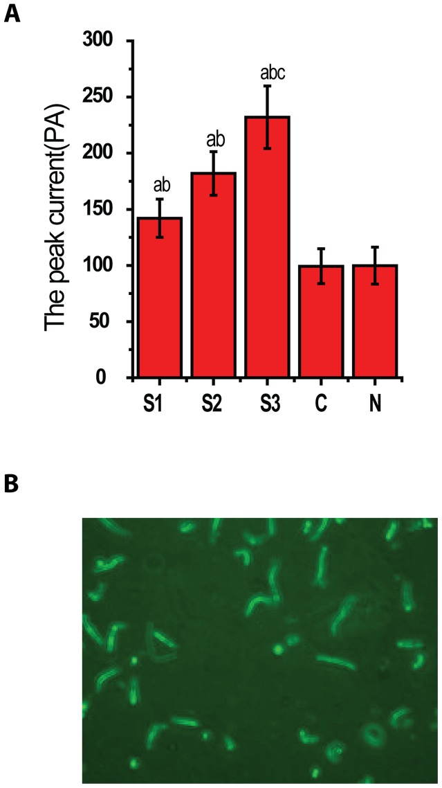
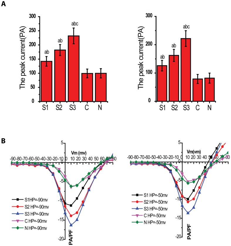
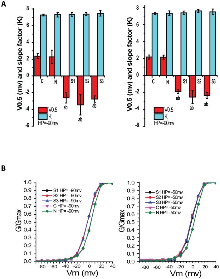
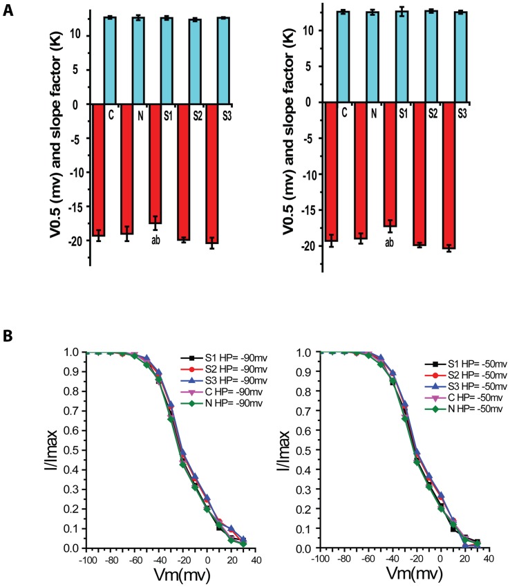
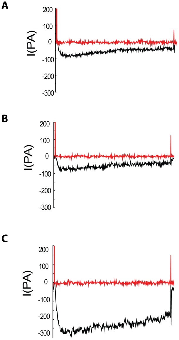
Methods: New Zealand white rabbits were randomly divided into five groups: sham (C), normal (N), 24 hours (S1), 48 hours (S2) and 72 hours (S3) after SAH. Non-heparinized autologous arterial blood (1 ml/kg) was injected into the cisterna magna to create SAH after intravenous anesthesia, and 1 ml/kg of saline was injected into cisterna magna in the sham group. Rabbits in group N received no injections. Basilar artery in S1, S2, S3 group were isolated at 24, 48, 72 hours after SAH. Basilar artery in group C was isolated at 72 hours after physiological saline injection. Basilar artery smooth muscle cells were isolated for all groups. Whole-cell patch-clamp technique was utilized to record cell membrane capacitance and VDCCs currents. The VDCCs antagonist nifedipine was added to the bath solution to block the Ca(++) channels currents.
Results: There were no significant differences in the number of cells isolated, the cell size and membrane capacitance among all the five groups. VDCC currents in the S1-S3 groups had higher amplitudes than those in control and sham groups. The significant change of current amplitude was observed at 72 hours after SAH, which was higher than those of 24 and 48 hours. The VDCCs were shown to expression in human artery smooth muscle cells.
Conclusions: The changes of activation characteristics and voltage-current relationship at 72 hours after SAH might be an important event which leads to a series of molecular events in the microenvironment of the basilar artery smooth muscle cells. This may be the key time point for potential therapeutic intervention against subarachnoid hemorrhage.
Conflict of interest statement
Figures
References
-
- Stornelli SA, French JD (1964) Subarachnoid Hemorrhage–Factors in Prognosis and Management. J Neurosurg 21: 769–780. - PubMed
-
- Hop JW, Rinkel GJ, Algra A, van Gijn J (1997) Case–fatality rates and functional outcome after subarachnoid hemorrhage: a systematic review. Stroke 28: 660–664. - PubMed
-
- Ishiguro M, Murakami K, Link T, Zvarova K, Tranmer BI, et al... (2008) Acute and chronic effects of oxyhemoglobin on voltage-dependent ion channels in cerebral arteries. Acta Neurochir Suppl 104: 99–102. - PubMed
-
- Wellman GC (2006) Ion channels and calcium signaling in cerebral arteries following subarachnoid hemorrhage. Neurol Res 28: 690–702. - PubMed
Publication types
MeSH terms
Substances
LinkOut - more resources
Full Text Sources
Other Literature Sources
