. 2014 Jan 2;9(1):e84740.
doi: 10.1371/journal.pone.0084740. eCollection 2014.

Neuromuscular fatigue is not different between constant and variable frequency stimulation

Affiliations

Neuromuscular fatigue is not different between constant and variable frequency stimulation

Maria Papaiordanidou et al. PLoS One. .

Abstract

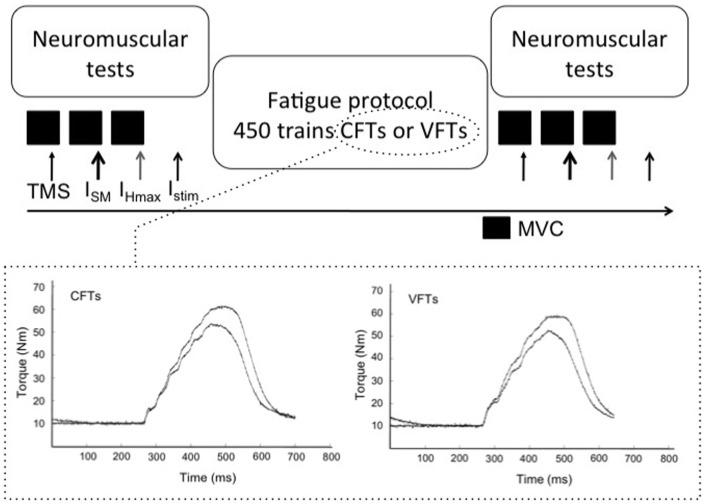
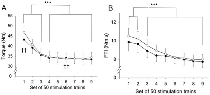
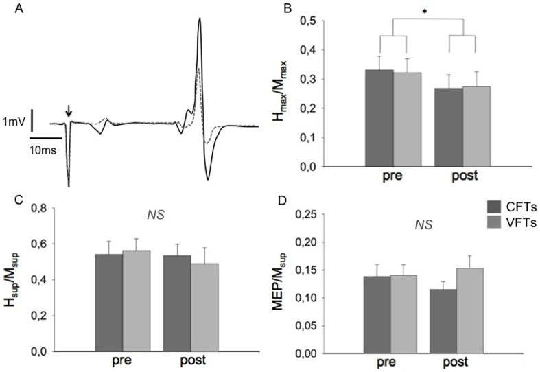
This study compared fatigue development of the triceps surae induced by two electrical stimulation protocols composed of constant and variable frequency trains (CFTs, VFTs, 450 trains, 30 Hz, 167 ms ON, 500 ms OFF and 146 ms ON, 500 ms OFF respectively). For the VFTs protocol a doublet (100 Hz) was used at the beginning of each train. The intensity used evoked 30% of a maximal voluntary contraction (MVC) and was defined using CFTs. Neuromuscular tests were performed before and after each protocol. Changes in excitation-contraction coupling were assessed by analysing the M-wave [at rest (Mmax) and during MVC (Msup)] and associated peak twitch (Pt). H-reflex [at rest (Hmax) and during MVC (Hsup)] and the motor evoked potential (MEP) during MVC were studied to assess spinal and corticospinal excitability of the soleus muscle. MVC decrease was similar between the protocols (-8%, P<0.05). Mmax, Msup and Pt decreased after both protocols (P<0.01). Hmax/Mmax was decreased (P<0.05), whereas Hsup/Msup and MEP/Msup remained unchanged after both protocols. The results indicate that CFTs and VFTs gave rise to equivalent neuromuscular fatigue. This fatigue resulted from alterations taking place at the muscular level. The finding that cortical and spinal excitability remained unchanged during MVC indicates that spinal and/or supraspinal mechanisms were activated to compensate for the loss of spinal excitability at rest.


Conflict of interest statement

Competing Interests: The authors have declared that no competing interests exist.

Figures

Figure 1
Figure 1. Schematic representation of the experimental protocol.
Neuromuscular tests were performed before and after the fatiguing protocols, which were composed of 450 trains of 6 pulses (CFTs or VFTs according to the session). The difference between protocols was that stimulation trains for the VFTs protocol started with an initial doublet (100 Hz). The lower panel shows the torque response of a representative subject on the first and last train of stimulation, for the CFTs (left graph) and VFTs (right graph) protocols. During the neuromuscular tests, superimposed and at rest stimulations (indicated with arrows) were delivered in a random order. TMS: transcranial magnetic stimulation, ISM: stimulation at supramaximal intensity, IHmax: stimulation at the intensity where Hmax was observed, Istim: stimulation at the intensity used during the fatiguing protocol, MVC: maximal voluntary contraction.
Figure 2
Figure 2. Characteristics of the fatiguing protocols.
White circles correspond to VFTs (variable frequency trains) and black circles represent CFTs (constant frequency trains). A. Torque time-course during the nine 50-train bouts. Torque significantly decreased for both protocols from the second 50-train set. Torque evoked during the first 50-train bout was significantly higher for VFTs. B. FTI (force-time integral) evolution during the protocols. FTI significantly decreased throughout both fatiguing protocols. ††P<0.01 significant difference between protocols, and ***P<0.001 significantly different from first bout values. Values are presented as means ± SEM.
Figure 3
Figure 3. Typical traces of the twitch elicited at the CFTs intensity from a representative subject.
Solid black lines correspond to recordings obtained before the protocol and dashed grey lines show the post-exercise recording. The upper panel depicts the mechanical response, and the lower one presents the electrophysiological responses. Note that for this subject, stimulation intensity evoked M-wave and H-reflex responses. Both mechanical and electrophysiological responses decreased at the post-stimulation stage.
Figure 4
Figure 4. Electrophysiological responses at pre- and post-fatigue conditions.
A. Typical recording of the Hmax response of a representative subject. Hmax was elicited at rest and was accompanied by a submaximal M-wave. The arrow indicates time of stimulation. The solid black line corresponds to pre-fatigue and the dashed grey line represents post-fatigue measurement. B, C and D show ratios (mean ± SEM) of Hmax/Mmax, Hsup/Msup and MEP/Msup respectively, obtained before and after the stimulation protocols. Dark grey bars represent the CFTs protocol while grey bars the VFTs protocol. * P<0.05 significant pre-post effect, NS not statistically significant.

References

    1. Hortobagyi T, Maffiuletti NA (2011) Neural adaptations to electrical stimulation strength training. Eur J Appl Physiol 111: 2439–2449. - PMC - PubMed
    1. Theurel J, Lepers R, Pardon L, Maffiuletti NA (2007) Differences in cardiorespiratory and neuromuscular responses between voluntary and stimulated contractions of the quadriceps femoris muscle. Respir Physiol Neurobiol 157: 341–347. - PubMed
    1. Gregory CM, Bickel CS (2005) Recruitment patterns in human skeletal muscle during electrical stimulation. Physical Therapy 85: 358–364. - PubMed
    1. Vanderthommen M, Duteil S, Wary C, Raynaud JS, Leroy-Willing A, et al. (2003) A comparison of voluntary and electrically induced contractions by interleaved 1H-and 31P-NMRS in humans. J Appl Physiol 94: 1012–1024. - PubMed
    1. Gerrits HL, De Haan A, Hopman MT, Van der Woude LHV, Jones DA, et al. (1999) Contractile properties of the quadriceps muscle in individuals with spinal cord injury. Muscle Nerve 22: 1249–1256. - PubMed

Publication types

LinkOut - more resources