

A Dynamic Dosimetry System for Prostate Brachytherapy
- PMID: 24392207
- PMCID: PMC3877329
- DOI: 10.1117/12.2008097
A Dynamic Dosimetry System for Prostate Brachytherapy
Abstract
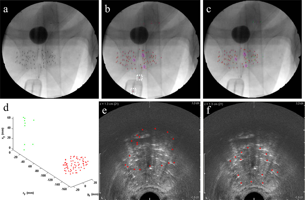
The lack of dynamic dosimetry tools for permanent prostate brachytherapy causes otherwise avoidable problems in prostate cancer patient care. The goal of this work is to satisfy this need in a readily adoptable manner. Using the ubiquitous ultrasound scanner and mobile non-isocentric C-arm, we show that dynamic dosimetry is now possible with only the addition of an arbitrarily configured marker-based fiducial. Not only is the system easily configured from accessible hardware, but it is also simple and convenient, requiring little training from technicians. Furthermore, the proposed system is built upon robust algorithms of seed segmentation, fiducial detection, seed reconstruction, and image registration. All individual steps of the pipeline have been thoroughly tested, and the system as a whole has been validated on a study of 25 patients. The system has shown excellent results of accurately computing dose, and does so with minimal manual intervention, therefore showing promise for widespread adoption of dynamic dosimetry.
Keywords: dynamic dosimetry; mobile non-isocentric C-arm; prostate brachytherapy; transrectal ultrasound.
Figures


Similar articles
-
Intraoperative Registered Ultrasound and Fluoroscopy (iRUF) for dose calculation during prostate brachytherapy: Improved accuracy compared to standard ultrasound-based dosimetry.Radiother Oncol. 2017 Jul;124(1):61-67. doi: 10.1016/j.radonc.2017.05.018. Epub 2017 Jun 21. Radiother Oncol. 2017. PMID: 28647400 Free PMC article.
-
Intra-operative 3D guidance in prostate brachytherapy using a non-isocentric C-arm.Med Image Comput Comput Assist Interv. 2007;10(Pt 2):9-17. doi: 10.1007/978-3-540-75759-7_2. Med Image Comput Comput Assist Interv. 2007. PMID: 18044547
-
Automatic segmentation of seeds and fluoroscope tracking (FTRAC) fiducial in prostate brachytherapy x-ray images.Proc SPIE Int Soc Opt Eng. 2010 Feb 23;7625:76252T. doi: 10.1117/12.844520. Proc SPIE Int Soc Opt Eng. 2010. PMID: 22977294 Free PMC article.
-
Does brachytherapy have a role in the treatment of prostate cancer?Hematol Oncol Clin North Am. 1996 Jun;10(3):653-73. doi: 10.1016/s0889-8588(05)70359-2. Hematol Oncol Clin North Am. 1996. PMID: 8773503 Review.
-
Place of modern imaging in brachytherapy planning.Cancer Radiother. 2018 Jun;22(4):326-333. doi: 10.1016/j.canrad.2018.03.005. Epub 2018 May 30. Cancer Radiother. 2018. PMID: 29858136 Review.
References
-
- Siegel R, Naishadham D, Jemal A. Cancer statistics, 2012. 1. Vol. 62. CA: A Cancer Jnl for Clinicians; 2012. pp. 10–29. - PubMed
-
- Nag S, Ciezki JP, Cormack R, Doggett S, DeWyngaert K, Edmundson GK, Stock RG, Stone NN, Yu Y, Zelefsky MJ. Intraoperative planning and evaluation of permanent prostate brachytherapy: Report of the american brachytherapy society. Intl. Jnl. of Radiation Oncology Biology Physics. 2001;51(5):1422–1430. - PubMed
-
- Jain A, Mustafa T, Zhou Y, Burdette C, Chirikjain GS, Fichtinger G. FTRAC – a robust fluoroscope tracking fiducial. Medical Physics. 2005;32(10):3185–3198. - PubMed
-
- Jain AK. PhD Thesis. JHU; 2008. Computation of 3D Implant Coordinates for Prostate Brachytherapy.
Grants and funding
LinkOut - more resources
Full Text Sources
Other Literature Sources