

Acetylcholinesterase function in apoptotic retina pigment epithelial cells induced by H2O2
- PMID: 24392323
- PMCID: PMC3874514
- DOI: 10.3980/j.issn.2222-3959.2013.06.06
Acetylcholinesterase function in apoptotic retina pigment epithelial cells induced by H2O2
Abstract
Aim: To investigate the acetylcholinesterase (AChE) expression involved in retina pigment epithelial (RPE) apoptosis induced by higher concentrations H2O2.
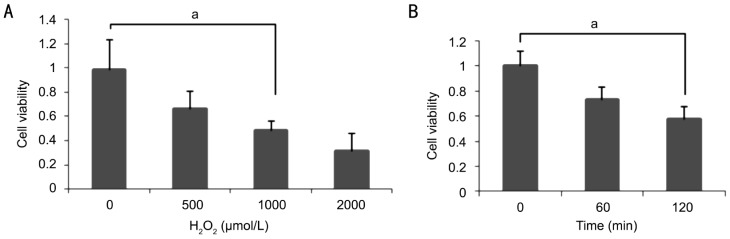
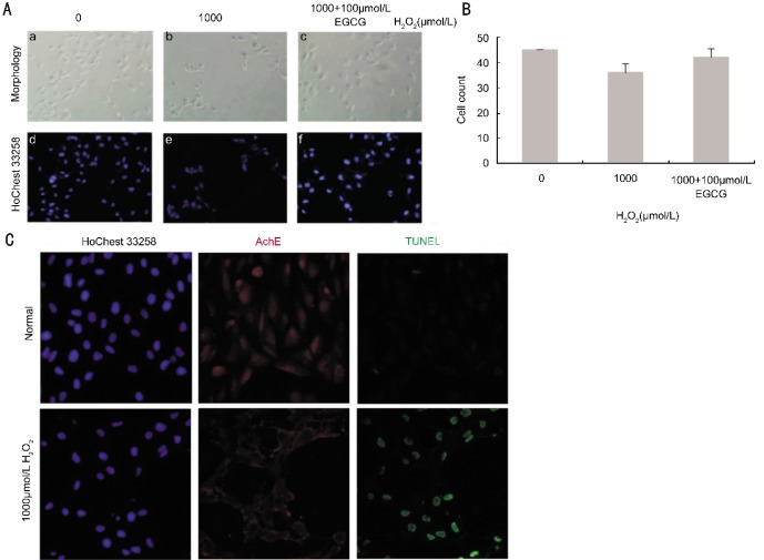
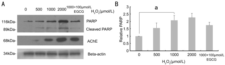
Methods: The human retinal pigment epithelium cell line ARPE-19 was from ATCC (Rockville, MD). Cultured ARPE-19 cells were treated with H2O2 at 0, 250, 500, 1 000, 2 000µmol/L and cell viability was measured with MTT assay. AChE expression and DNA fragments were analyzed by immunocytochemistry, TUNEL and PARP-1 Western blotting.
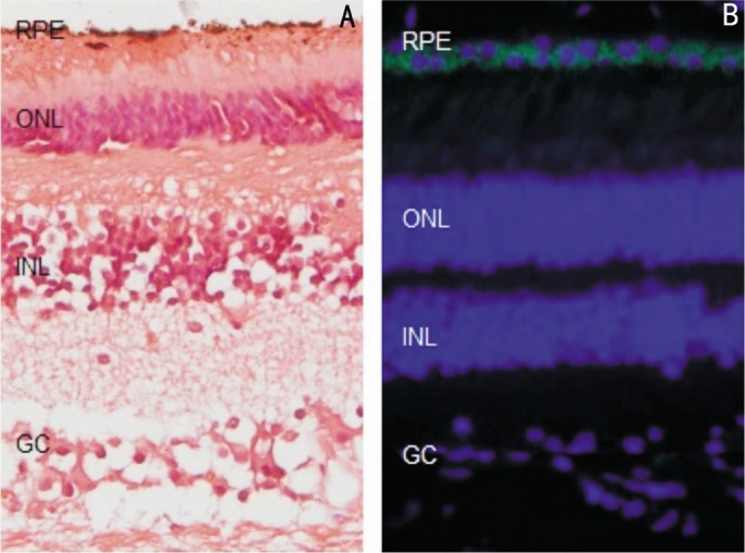
Results: Immunofluorescence detected AChE exist in the normal human retinal tissue. When H2O2 >500µmol/L, AChE expression showed an increase after 2h, and this concentration was selected for the present study. RPE cell was induced with 1 000µmol/L H2O2 for 2h, compared to the control group, cell activity decline detected by MTT, AChE and PARP-1 protein expression was significantly increased detected by Western blotting. AChE immunofluorescence staining was positive in RPE cell after H2O2 incubate 2h. In addition, pretreatment with 100µmol/L epigallocatechin gallate (EGCG), cell viability increased from 31.20%±3.90% to 70.23%±12.96%.
Conclusion: AChE is weakly expressed in normal human RPE cells. Stimulation with H2O2 caused the stable increase of AChE expression in RPE cells, which may indicate that AChE may be an important role in AMD.
Keywords: acetylcholinesterase; age-related macular degeneration; oxidative stress; retina pigment epithelial cells.
Figures
References
-
- Cheung CM, Tai ES, Kawasaki R, Tay WT, Lee JL, Hamzah H, Wong TY. Prevalence of and risk factors for age-related macular degeneration in a multiethnic Asian cohort. Arch Ophthalmol. 2012;130(4):480–486. - PubMed
-
- Klein R, Chou CF, Klein BE, Zhang X, Meuer SM, Saaddine JB. Prevalence of age-related macular degeneration in the US population. Arch Ophthalmol. 2011;129(1):75–80. - PubMed
-
- Minassian DC, Reidy A, Lightstone A, Desai P. Modelling the prevalence of age-related macular degeneration (2010-2020) in the UK: expected impact of anti-vascular endothelial growth factor (VEGF) therapy. Br J Ophthalmol. 2011;95(10):1433–1436. - PubMed
-
- Cai X, McGinnis JF. Oxidative stress: the achilles' heel of neurodegenerative diseases of the retina. Front Biosci. 2012;17:1976–1995. - PubMed
LinkOut - more resources
Full Text Sources
Other Literature Sources
Miscellaneous