Review
. 2014:54:537-58.
doi: 10.1146/annurev-pharmtox-011112-140338.

Turning off AKT: PHLPP as a drug target

Affiliations
Review

Turning off AKT: PHLPP as a drug target

Alexandra C Newton et al. Annu Rev Pharmacol Toxicol. 2014.

Abstract

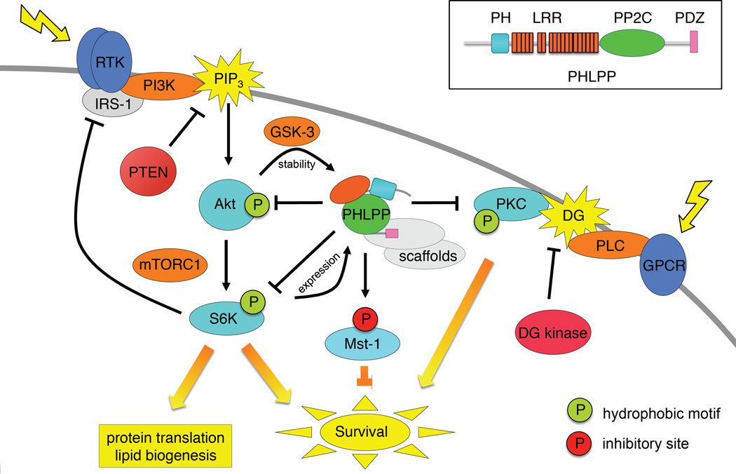
Precise control of the balance between protein phosphorylation, catalyzed by protein kinases, and protein dephosphorylation, catalyzed by protein phosphatases, is essential for cellular homeostasis. Dysregulation of this balance leads to pathophysiological states, driving diseases such as cancer, heart disease, and diabetes. Aberrant phosphorylation of components of the pathways that control cell growth and cell survival are particularly prevalent in cancer. One of the most studied tumor suppressors in these pathways is the lipid phosphatase PTEN (phosphatase and tensin homolog deleted on chromosome ten), which dephosphorylates the lipid second messenger phosphatidylinositol 3,4,5-trisphosphate (PIP3), thus preventing activation of the oncogenic kinase AKT (v-akt murine thymoma viral oncogene homolog). In 2005, the discovery of a family of protein phosphatases whose members directly dephosphorylate and inactivate AKT introduced a new negative regulator of the phosphoinositide 3-kinase (PI3K) oncogenic pathway. Pleckstrin homology domain leucine-rich repeat protein phosphatase (PHLPP) isozymes comprise a novel tumor suppressor family whose two members, PHLPP1 and PHLPP2, are deleted as frequently as PTEN in cancers such as those of the prostate. PHLPP is thus a novel therapeutic target to suppress oncogenic pathways and is a potential candidate biomarker to stratify patients for the appropriate targeted therapeutics. This review discusses the role of PHLPP in terminating AKT signaling and how pharmacological intervention would impact this pathway.


Figures

Figure 1
Figure 1. AKT is a central mediator of PI3K signaling
Cartoon showing details of the regulation of AKT: AKT is co-translationally phosphorylated on the turn motif (orange circle, Thr450 in AKT1) by ribosome and ER-localized mTORC2 (orange oval). This phosphorylation stabilizes AKT and protects it from proteosome-mediated degradation. Signals that generate PIP3 engage AKT’s PH domain and thus recruit it to the plasma membrane (middle panel; membrane-engaged species). Membrane-binding unmasks the activation loop, resulting in phosphorylation by PDK-1 (pink circle, Thr308 in AKT1) and a subsequent tightly-coupled phosphorylation on the hydrophobic motif (green circle, Ser473 in AKT1). Although mTORC2 has been proposed to phosphorylate this site too, mechanisms that displace the PH domain (e.g. tethering AKT to the membrane via myristoylation) bypass the requirement for mTORC2. This latter phosphorylation depends on the intrinsic catalytic activity of AKT, suggesting that, similar to PKC, this site is autophosphorylated following structuring of the active site by the PDK-1 phosphorylation of Thr308. The fully-phosphorylated species of AKT is locked in an active conformation and diffuses throughout the cell to mediate down stream signaling (bottom right species). Signaling is suppressed by dephosphorylation of PIP3 by the tumor suppressor PTEN and acutely terminated by direct dephosphorylation of AKT to regenerate the mono-phosphorylated (phospho-Thr 450), auto-inhibited species. Dephosphorylation is catalyzed in part by the recently discovered PHLPP phosphatases, which directly dephosphorylate the hydrophobic motif, and by okadaic acid-sensitive phosphatases, such as PP2A, which dephosphorylate the activation loop.
Figure 2
Figure 2. PHLPP suppresses lipid second messenger signaling
PHLPP (center) directly dephosphorylates and inactivates AKT, PKC, and S6K, at their hydrophobic phosphorylation motif (green circle) thus opposing cell survival and growth signals, and directly dephosphorylates and activates Mst1 at an inhibitory site (red circle), thus promoting pro-apoptotic signals. Indicated are two negative feedback loops which control the steady-state levels of PHLPP: high AKT activity up regulates PHLPP levels by suppressing GSK-3-dependent degradation, and high S6K activity up regulates PHLPP expression. Note that targeting the PI3 kinase pathway within these feedback loops will have the consequence of decreasing PHLPP levels, thus promoting pro-survival pathways mediated by PKC and inhibiting pro-apoptotic pathways mediated by Mst1. The lipid phosphatase PTEN opposes the lipid kinase phosphatidylinositol 3 kinase (PI3K) by dephosphorylating PIP3 and the lipid kinase DG kinase opposes phospholipase C by phosphorylating DG. Scaffolds play an essential role in controlling substrate specificity of PHLPP and are indicated in grey. Also shown is the negative feedback loop from S6K to the scaffold IRS-1, which dampens PI3K activation and hence AKT activation (see Figure 4 also). Inset shows conserved domain structure of mammalian PHLPP family members showing PH domain (cyan), Leucine-rich repeats (LRR; orange), PP2C domain (green), and C-terminal PDZ-binding motif (pink); PHLPP1β and PHLPP2 also have a predicted RA domain preceding the PH domain (not shown).
Figure 3
Figure 3. PHLPP phosphatase domain structure and model with hydrophobic motif peptide substrate bound
A. Homology model of PP2C domain of PHLPP2, based on crystal structure of PP2Cα (47), showing two of the active site acidic residues (Asp 806, Asp 1024; pink) that coordinate two Mn2+ (yellow spheres) (48); Leu 1016 which is present as Ser in 30% of the population is highlighted in orange. B. Surface rendition of the active site of PHLPP2 docked with a phosphorylated hydrophobic motif peptide from AKT (HFPQFpSYSAS; phosphorylated Ser (Ser473) in magenta; active site residues and Mn2+ colored as in A.
Figure 4
Figure 4. Feedback loops and therapy
mTORC1 activation in cancer results in increased protein translation needed for cell growth and proliferation (black arrows). When mTORC1 signaling is strong, mTORC1 activates three tumor suppressive responses (red arrows) that attenuate the pathway via 1] increasing PHLPP or 2] irreversibly arrest the cell by increasing p53. In prostate, androgen receptor (AR) is activated by RTK signaling and limits AKT signaling through FKBP5-mediated increase in PHLPP activity towards AKT. The pathway’s tumor suppressive feedbacks can be inadvertently blocked by therapeutics (green arrows). First, AR blockade by hormone therapy inhibits PHLPP suppression of AKT, thus activating AKT. Second, mTORC1-targeting kinase inhibitors such as rapalogs block three known negative feedbacks: S6 kinase-dependent negative feedback loop to PI3K signaling via RTKs, mTORC1-dependent increase in PHLPP levels, and mTORC1-dependent increases in p53 levels.

References

    1. Taylor BS, Schultz N, Hieronymus H, Gopalan A, Xiao Y, et al. Integrative genomic profiling of human prostate cancer. Cancer Cell. 18:11–22. - PMC - PubMed
    1. Gao J, Aksoy BA, Dogrusoz U, Dresdner G, Gross B, et al. Integrative Analysis of Complex Cancer Genomics and Clinical Profiles Using the cBioPortal. Sci Signal. 2013;6:pl1. - PMC - PubMed
    1. Vanhaesebroeck B, Leevers SJ, Ahmadi K, Timms J, Katso R, et al. Synthesis and function of 3-phosphorylated inositol lipids. Annu Rev Biochem. 2001;70:535–602. - PubMed
    1. Toker A. Protein kinases as mediators of phosphoinositide 3-kinase signaling. Mol Pharmacol. 2000;57:652–658. - PubMed
    1. Chan TO, Rittenhouse SE, Tsichlis PN. AKT/PKB and other D3 phosphoinositide-regulated kinases: kinase activation by phosphoinositide-dependent phosphorylation. Annu Rev Biochem. 1999;68:965–1014. - PubMed

Publication types

MeSH terms