. 2014 Jan 6:12:4.
doi: 10.1186/1479-5876-12-4.

Ovarian cancer cell invasiveness is associated with discordant exosomal sequestration of Let-7 miRNA and miR-200

Affiliations

Ovarian cancer cell invasiveness is associated with discordant exosomal sequestration of Let-7 miRNA and miR-200

Miharu Kobayashi et al. J Transl Med. .

Abstract

Background: The role of exosomes in the pathogenesis and metastatic spread of cancer remains to be fully elucidated. Recent studies support the hypothesis that the release of exosomes from cells modifies local extracellular conditions to promote cell growth and neovascularisation. In addition, exosomes may modify the phenotype of parent and/or target cell. For example, sequestration of signaling mediators into exosomes may reduce their intracellular bioavailability to the parent cell thereby altering cell phenotype and metastatic potential. The fusion of released exosomes with target cell and delivery may also modify cell function and activity. In this study, to further elucidate the role of exosomes in ovarian cancer, the release of exosomes from two ovarian cancer cell lines of different invasive capacity and their miRNA content of exosomes were compared. The hypothesis to be tested was that ovarian cancer cell invasiveness is associated with altered release of exosomes and discordant exosomal sequestration of miRNA.

Methods: High (SKOV-3) and low (OVCAR-3) invasive ovarian cancer cell lines were used to characterize their exosome release. SKOV-3 and OVCAR-3 cells were cultured (DMEM, 20% exosome-free FBS) under an atmosphere of 8% O2 for 24 hours. Cell-conditioned media were collected and exosomes were isolated by differential and buoyant density centrifugation and characterised by Western blot (CD63 and CD9). Exosomal microRNA (let-7a-f and miR-200a-c) content was established by real-time PCR.

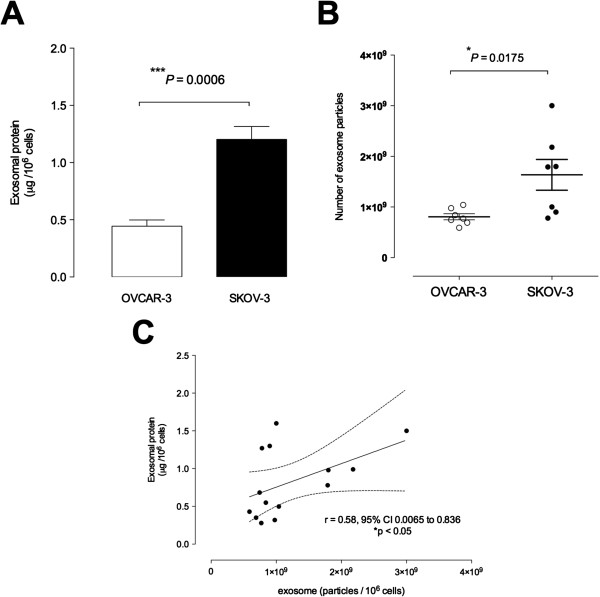
Results: Exosomes were identified with by the presence of typical cup-shaped spherical vesicle and the expression of exosome markers: CD63, CD9. SKOV-3 cells released 2.7-fold more exosomes (1.22 ± 0.11 μg/106 cells) compared to OVCAR-3 (0.44 ± 0.05 μg/106 cells). The let-7 family miRNA transcripts were identified in both ovarian cancer cell lines and their exosomes. The let-7 family transcripts were more abundant in OVCAR-3 cell than SKOV-3 cells. In contrast, let-7 family transcripts were more abundant in exosomes from SKOV-3 than OVCAR-3. miR-200 family transcripts were only identified in OVCAR-3 cells and their exosomes.

Conclusions: The data obtained in this study are consistent with the hypothesis that the releases of exosomes varies significantly between ovarian cancer cell lines and correlates with their invasive potential.


Figures

Figure 1
Figure 1
Ovarian cancer cell invasion. OVCAR-3 and SKOV-3 cells were grown to confluence in complete media. A wound was made using 96 well WoundMaker and then overlaid to form a 3D matrix-gel. Invasion assay was preformed with OVCAR-3 and SKOV-3 cells in complete media. Cells were imaged with IncuCyte™ (Essen BioSciences, USA) every three hours for 24 h. (see Methods). (A) Representative images of OVCAR-3 (A1-A4) and SKOV-3 (A5-A8) cell invasion. A1 and A5: phase contrast micrograph image immediately after wounding; A2 and A6: graphical representation showing the calculation of initial wound width; A3 and A7: cells images show 24 hours later; A4 and A8: graphical representation of cell invasion at 24 hours of the experiment. The grey region denotes the area of the initial wound covered by advancing cells. (B) The time course of OVCAR-3 and SKOV-3 invasion. (C) Area under curves analysis from B. Data represented as mean ± SEM (n = 6). In C, *P < 0.001 versus SKOV-3.
Figure 2
Figure 2
Characterisation of exosomes from ovarian cancer cell lines. (A) Representative image of western blot for the presence of CD63 and CD9. S1-S4 and S5-S6 represent different exosomes isolation from OVCAR-3 and SKOV-3 cells, respectively. (B) Representative electron micrograph of exosomes isolated from OVCAR-3 and SKOV-3 cells. (C) Nanosight measurement of particle-size distribution in preparation from OVAR-3 (black) and SKOV-3 (red) exosomes (see Methods). In B, Scale bar 100 nm.
Figure 3
Figure 3
Exosome releases from ovarian cancer cell lines. Exosomes were quantified in culture media of ovarian cancer cell lines using an ELISA kit (see Methods). (A) Levels of exosome as presented as protein concentration from1x106 OVCAR-3 (white bar) and SKOV-3 cells (black bar). (B) Quantification of number of exosome particles from OVCAR-3 (white circles) and SKOV-3 cells (black circles) per 106 cells. (C) Relationship between number of exosome particles and exosomal protein concentration from OVCAR-3 and SKOV-3 cells. Fitted linear regression line with 95% confidence intervals (dotted lines). In A, ***P < 0.01 versus SKOV-3. In B, *P < 0.05 versus SKOV-3.
Figure 4
Figure 4
Expression of let-7 and miR-200 families. Total microRNA was extracted using the Ambion mirVana PARIS Kit (Invitrogen). Real-time PCR was performed with miScript SYBR Green Kit to compare let-7 and miR-200 family expression between ovarian cancer cell lines (OVAR-3 and SKOV-3 cells) (A) and between cell-derived exosomes (exo-OVAR-3 and exo-SKOV-3) (B). Data are represented as mean ± SEM (n = 6). In A and B, ANOVA p < 0.0001 between groups. In B, *p < 0.001 vs exo-SKOV-3.
Figure 5
Figure 5
Comparison of miRNA transcripts expression between cells and exosomes. The miRNA expressions between cells and exosomes in OVCAR-3 (A) and SKOV-3 (B). Data is represented by mean ± SEM (n = 6 per group).
Figure 6
Figure 6
Proposed model of ovarian cancer cell-derived exosome action. Tumour cells with different invasion capacity exist in a single epithelial ovarian tumour, releasing exosomes. High invasive tumour cells (SKOV-3) release significantly more exosomes (1), containing proteins involved in processes such as: cell death and survival, cellular movement, cancer, cell-to-cell signalling and interaction, cellular growth and proliferation. The let-7 family that are known to suppress cell proliferation was significantly more expressed in exosomes from high invasive exosomes (from SKOV-3). On the other hand, the miR-200 family that suppress epithelial to mesenchymal transition was only expressed in exosomes derived from low invasive cells (from OVCAR-3) (2). The miRNA profile differs between exosomes derived from cells with different invasive capacity (OVAR-3 versus SKOV-3) (3).

Similar articles

Cited by

References

    1. Jemal A, Bray F, Center MM, Ferlay J, Ward E, Forman D. Global cancer statistics. CA Cancer J Clin. 2011;61:69–90. doi: 10.3322/caac.20107. - DOI - PubMed
    1. Siegel R, Naishadham D, Jemal A. Cancer statistics, 2012. CA Cancer J Clin. 2012;62:10–29. doi: 10.3322/caac.20138. - DOI - PubMed
    1. Jemal A, Siegel R, Ward E, Hao Y, Xu J, Thun MJ. Cancer statistics, 2009. CA Cancer J Clin. 2009;59:225–249. doi: 10.3322/caac.20006. - DOI - PubMed
    1. Taylor DD, Gercel-Taylor C. Tumour-derived exosomes and their role in cancer-associated T-cell signalling defects. Br J Cancer. 2005;92:305–311. - PMC - PubMed
    1. Simpson RJ, Jensen SS, Lim JW. Proteomic profiling of exosomes: current perspectives. Proteomics. 2008;8:4083–4099. doi: 10.1002/pmic.200800109. - DOI - PubMed

Publication types