. 2014 Jan 24;443(4):1280-5.
doi: 10.1016/j.bbrc.2013.12.135. Epub 2014 Jan 4.

Matrix rigidity regulates spatiotemporal dynamics of Cdc42 activity and vacuole formation kinetics of endothelial colony forming cells

Affiliations

Matrix rigidity regulates spatiotemporal dynamics of Cdc42 activity and vacuole formation kinetics of endothelial colony forming cells

Seung Joon Kim et al. Biochem Biophys Res Commun. .

Abstract

Recent evidence has shown that endothelial colony forming cells (ECFCs) may serve as a cell therapy for improving blood vessel formation in subjects with vascular injury, largely due to their robust vasculogenic potential. The Rho family GTPase Cdc42 is known to play a primary role in this vasculogenesis process, but little is known about how extracellular matrix (ECM) rigidity affects Cdc42 activity during the process. In this study, we addressed two questions: Does matrix rigidity affect Cdc42 activity in ECFC undergoing early vacuole formation? How is the spatiotemporal activation of Cdc42 related to ECFC vacuole formation? A fluorescence resonance energy transfer (FRET)-based Cdc42 biosensor was used to examine the effects of the rigidity of three-dimensional (3D) collagen matrices on spatiotemporal activity of Cdc42 in ECFCs. Collagen matrix stiffness was modulated by varying the collagen concentration and therefore fibril density. The results showed that soft (150 Pa) matrices induced an increased level of Cdc42 activity compared to stiff (1 kPa) matrices. Time-course imaging and colocalization analysis of Cdc42 activity and vacuole formation revealed that Cdc42 activity was colocalized to the periphery of cytoplasmic vacuoles. Moreover, soft matrices generated faster and larger vacuoles than stiff matrices. The matrix-driven vacuole formation was enhanced by a constitutively active Cdc42 mutant, but significantly inhibited by a dominant-negative Cdc42 mutant. Collectively, the results suggest that matrix rigidity is a strong regulator of Cdc42 activity and vacuole formation kinetics, and that enhanced activity of Cdc42 is an important step in early vacuole formation in ECFCs.

Keywords: Endothelial colony forming cells (ECFCs); Fluorescence resonance energy transfer (FRET); Live cell imaging; Matrix stiffness; Mechanotransduction; Rho family GTPases.


Figures

Fig. 1
Fig. 1
(A) Molecular structure of a FRET-based Cdc42 biosensor. Adapted from ref. [22]. (B) Schematic diagram showing timeline of ECFC transfection, collagen-based tissue construct formation, and spatiotemporal analysis of ECFC vacuole formation and Cdc42 activity.
Fig. 2
Fig. 2
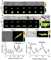
Colocalization of Cdc42 activation and vacuole formation. (A) Time-lapse images of early vacuole formation and Cdc42 activity (18 to 30 hours following seeding) for an individual ECFC within a (A) soft (150 Pa) and (B) stiff (1 kPa) collagen matrix. Color bars represent emission ratio of YFP/CFP of the biosensor, an index of Cdc42 activation. Close-up view of boxed area in (B) shows colocalization of regional Cdc42 activation with the vacuole periphery. Arrowheads indicate strong Cdc42 activation and vacuoles. (C, D) Cdc42 activity profile along the line from the left to the right on the cell body in a (C) soft and (D) stiff matrix. Scale bars, 10 μm.
Fig. 3
Fig. 3
Effect of matrix rigidity on the dynamics of Cdc42 activity and vacuole formation kinetics. (A) Cdc42 activation time course during early vacuole formation in soft and stiff matrices. 150 Pa: n=11 cells; 1 kPa; n=13 cells. (B) Basal levels of Cdc42 activity of ECFCs (150 Pa: n=7 cells; 1kPa: n=8 cells) grown in two different matrices before induction of vacuole formation. * p < 0.05. (C) Percentage of vacuole-forming cells as a function of time following vacuole induction in soft and stiff matrices. Data collected from >150 cells for 3 different experiments per group.
Fig. 4
Fig. 4
Role of matrix rigidity and Cdc42 in vacuole formation. (A) ECFCs form larger vacuoles in soft matrices than in stiff matrices (150Pa: n = 14 cells; 1kPa: n=16 cells). (B) Representative DIC images of ECFCs forming vacuoles in two different matrices. Scale bars, 10 μm. (C) The percentage of vacuole forming cells increases significantly with expression of Cdc42-L61 compared to untreated control. In contrast, expression of Cdc42-N17 yields a significant decrease in the percentage of vacuole-forming cells. Data collected from >200 cells for 5 different experiments per group. *p < 0.05, ** p < 0.01, *** p < 0.001.

References

    1. Critser PJ, Voytik-Harbin SL, Yoder MC. Isolating and defining cells to engineer human blood vessels. Cell Prolif. 2011;44(Suppl 1):15–21. - PMC - PubMed
    1. Ingram DA, Mead LE, Tanaka H, Meade V, Fenoglio A, Mortell K, Pollok K, Ferkowicz MJ, Gilley D, Yoder MC. Identification of a novel hierarchy of endothelial progenitor cells using human peripheral and umbilical cord blood. Blood. 2004;104:2752–2760. - PubMed
    1. Watt SM, Athanassopoulos A, Harris AL, Tsaknakis G. Human endothelial stem/progenitor cells, angiogenic factors and vascular repair. J R Soc Interface. 2010;7(Suppl 6):S731–751. - PMC - PubMed
    1. Critser PJ, Yoder MC. Endothelial colony-forming cell role in neoangiogenesis and tissue repair. Curr Opin Organ Transplant. 2010;15:68–72. - PMC - PubMed
    1. Bailey JL, Critser PJ, Whittington C, Kuske JL, Yoder MC, Voytik-Harbin SL. Collagen oligomers modulate physical and biological properties of three-dimensional self-assembled matrices. Biopolymers. 2011;95:77–93. - PMC - PubMed

Publication types

MeSH terms

LinkOut - more resources