. 2014:5:2914.
doi: 10.1038/ncomms3914.

The pseudogene TUSC2P promotes TUSC2 function by binding multiple microRNAs

Affiliations
Free PMC article

The pseudogene TUSC2P promotes TUSC2 function by binding multiple microRNAs

Zina Jeyapalan Rutnam et al. Nat Commun. 2014.
Free PMC article

Abstract

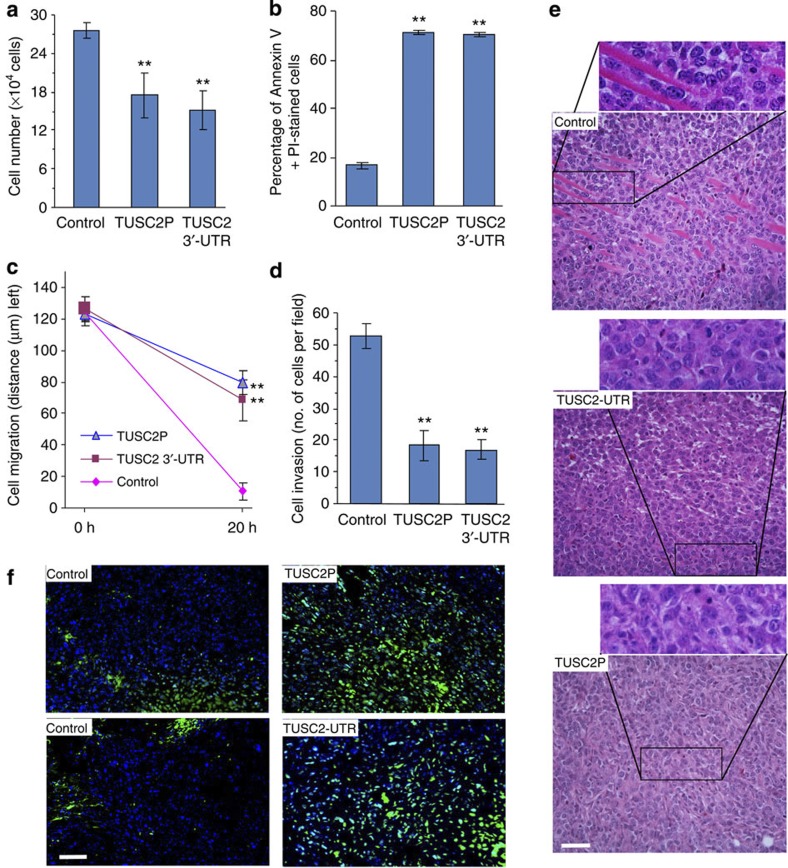
Various non-coding regions of the genome, once presumed to be 'junk' DNA, have recently been found to be transcriptionally active. In particular, pseudogenes are now known to have important biological roles. Here we report that transcripts of the two tumour suppressor candidate-2 pseudogenes (TUSC2P), found on chromosomes X and Y, are homologous to the 3'-UTR of their corresponding protein coding transcript, TUSC2. TUSC2P and the TUSC2 3'-UTR share many common miRNA-binding sites, including miR-17, miR-93, miR-299-3p, miR-520a, miR-608 and miR-661. We find that ectopic expression of TUSC2P and the TUSC2 3'-UTR inhibits cell proliferation, survival, migration, invasion and colony formation, and increases tumour cell death. By interacting with endogenous miRNAs, TUSC2P and TUSC2 3'-UTR arrest the functions of these miRNAs, resulting in increased translation of TUSC2. The TUSC2P and TUSC2 3'-UTR could thus be used as combinatorial miRNA inhibitors and might have clinical applications.


Figures

Figure 1
Figure 1. Expression of the TUSC2P pseudogene.
(a) Analysis of miRNA targeting the 3′-UTR of TUSC2 and pseudogene TUSC2P on chromosome Y and chromosome X. (b) Expression of TUSC2P was analysed in different cancer and non-cancer cell lines by real-time PCR. The cancer cell lines had a decreased amount of TUSC2P mRNA compared with the non-cancer cell lines. The inset represents bands of reverse transcription–PCR of these cell lines; n=4, error bar, s.d. (c) TUSC2P and TUSC2 3′-UTR were cloned and inserted into pcDNA3.1 plasmid, downstream of the cytomegalovirus promoter, producing the TUSC2P and TUSC2-UTR constructs. (d) Total RNA isolated from 4T1 cells stably transfected with TUSC2P, TUSC2-UTR and control vector were analysed by real-time PCR. The TUSC2P-transfected cells had a fourfold increase compared with the endogenous TUSC2P levels in the cells. The TUSC2-UTR-transfected cells had approximately a threefold increased expression of TUSC2 compared with the control cells; n=3, **P<0.01 analysed by t-test, error bar, s.d.
Figure 2
Figure 2. TUSC2P and TUSC2 3′-UTR can function as competing endogenous RNAs (ceRNAs).
(a) The TUSC2P, TUSC2-UTR and control 4T1 cells were maintained in tissue culture dishes in serum-free conditions. Surviving cells were collected and counted after 8 days. The TUSC2P and TUSC2-UTR cells had a decreased rate of survival; n=3, **P<0.01 analysed by t-test, error bar, s.d. (b) The cells were cultured in serum-free conditions for 5 days, followed by apoptosis analysis. After being stained with Annexin V and PI, the cells were analysed by flow cytometry. The TUSC2P- and TUSC2-UTR-transfected cells had larger populations of apoptotic and necrotic cells (Annexin V positive, PI positive and Annexin V, PI positive) compared with the control cells; n=3, **P<0.01 by t-test, error bar, s.d. (c) Migration rates were monitored using scratch assay. It was found that the TUSC2P- and TUSC2-UTR-transfected 4T1 cells had a slower rate of migration compared with the control cells. **P<0.01 by t-test, error, s.d., n=10. (d) Cell invasion was examined using Matrigel-coated transwell chambers. When cells were placed in the chambers, the TUSC2P and TUSC2-UTR cells travelled slower through the Matrigel and membrane pores, and spread out on the underside of the membrane significantly less than the control cells; n=5, **P<0.01 by t-test, error bar, s.d. (e) Haematoxylin and eosin staining was performed in tumours formed by injecting TUSC2-UTR, TUSC2P and control 4T1 cells into mice. Evidence of invasive tissues could be seen in the control tumours. However, it was absent in the TUSC2P and TUSC2-UTR tumours. Scale bar, 50 μm. (f) TdT-mediated dUTP nick end labelling staining was performed to observe the apoptotic cells in the tumour sections. Tumours formed by the TUSC2P- and TUSC2-UTR-transfected cells displayed an increased amount of apoptotic cells than the control. Scale bar, 50 μm.
Figure 3
Figure 3. Targeting of TUSC2 and TIMP2/3 by miRNAs.
(a) We hypothesized that the TUSC2P/TUSC2-UTR would attract endogenous miR-17, miR-93, miR-299-3p, miR-520a, miR-608 and miR-661, thus freeing TUSC2, TIMP2 and TIMP3 mRNAs to be available for translation. (b) Computation analysis of the TUSC2 showed potential binding sites for miR-93, miR-299-3p, miR-520a, miR-17, miR-608 and miR-661. (c) Computation analysis of the TUSC2P showed potential binding sites for miR-93, miR-17, miR-520a, miR-608 and miR-661. (d) Computation analysis of TIMP2 3′-UTR (upper) and TIMP3 (lower) 3′-UTRs revealed binding sites for miR-17, miR-93, miR-299-3p, miR-608 and miR-661. (e) Protein lysates were prepared from TUSC2P-, TUSC2-UTR- and vector-transfected cells and subjected to western blot analysis probed with anti-TUSC2, anti-TIMP2 and anti-TIMP3 antibodies. There was elevated expression of TUSC2, TIMP2 and TIMP3 in the TUSC2P and TUSC2-UTR cells compared with the control cells. Detection of β-actin on the same membranes served as a loading control. (f) Protein lysates were prepared from TUSC2P and control tumours, and subject to western blot analysis and probed with anti-TUSC2, anti-TIMP2 and anti-TIMP3 antibodies. TUSC2, TIMP2 and TIMP3 were upregulated in the TUSC2P tumours.
Figure 4
Figure 4. Expression of TUSC2 and TIMP2/3 in different cell lines and patient specimens.
(ac) Expression of TUSC2 (a), TIMP2 (b) and TIMP3 (c) were analysed by real-time PCR for mRNA levels and western blotting for protein levels (insets) in different cell lines as indicated; n=4, error bar, s.d. (d) Human breast cancer specimens were probed for expression of TUSC2, TIMP2 and TIMP3. Expression of these molecules was detected in the ductal structures (arrows), but not in the tumour tissues. Scale bar, 50 μm.
Figure 5
Figure 5. Confirmation of TIMP2 and TIMP3 targeting.
(af) Fragments of TUSC2P harbouring different target sites of miR-299-3p (a), miR-661 (b), miR-17 (c), miR-93 (d), miR-520a (e) and miR-608 (f) were cloned into the luciferase reporter vector pMir-Report. 4T1 cells were co-transfected with one of these miRNAs and the luciferase constructs or a mutant constructs, in which the miRNA target sites were mutated. A non-related fragment of complementary DNA was used as a positive control. Luciferase activity assays indicated that all six miRNAs repressed luciferase activities when it harboured the corresponding TUSC2P fragment, which was reversed when the potential miRNA target site was mutated; n=3, **P<0.01 (between wild type and mutants) by t-test, error bar, s.d. (gk) The cells were co-transfected with luciferase reporter plasmids for TIMP2 with corresponding miRNAs or a control oligo. In the presence of the targeting UTR, luciferase activities were inhibited, which were significantly elevated when the target sites were mutated; n=3, **P<0.01 (between wild type and mutants) by t-test, error bar, s.d. (lo) The TIMP3 3′-UTR fragments containing the binding sites for miR-17, miR-93, miR-299-3p and miR-661 were cloned into pMir-Report. Luciferase activity assays indicated that all four miRNAs repressed luciferase activities, which was reversed when the miRNA binding site was mutated; n=3, **P<0.01 (between wild type and mutants) by t-test, error bar, s.d.
Figure 6
Figure 6. Functional analysis of miRNAs targeting TUSC2.
(a) Lysates of 4T1 cells transfected with miRNAs were analysed by western blotting. Expression of miR-17, miR-93, miR-299-3p, miR-520a, miR-608 and miR-661 decreased TUSC2 protein levels. β-Actin on the same membranes served as a loading control. (b) The cells transfected with different miRNAs were subject to western blot analysis for expression of TIMP2 and TIMP3. Both TIMP2 and TIMP3 levels decreased in cells transfected with miRNAs as compared with the control oligo. (c) Apoptosis was analysed in cells transfected with different miRNAs. Transfection with the miRNAs decreased apoptosis; n=3, **P<0.01. (d) The migration rate was measured for the miRNA-transfected 4T1 cells. There was an increased amount of cell migration in miRNA-transfected cells compared with control cells; n=10, **P<0.01 by t-test, error bar, s.d. (e) In cell invasion assays, the cells transfected with miR-17, miR-93, miR-299-3p, miR-520a, miR-608 and miR-661 showed drastically increased in the capacity of invasion through the membrane pores compared with the control cells; n=5, **P<0.01 by t-test.
Figure 7
Figure 7. Expression of TUSC2P affects activities of TUSC2 and TIMP3.
(a,b) Copy number of TUSC2 and TUSC2P was determined in vector- and TUSC2P-transfected cells. P-values are obtained by t-test and shown in the figure; n=3, error bar, s.d. (c) Copy number of TUSC2P was determined in vector- and TUSC2P-transfected cells. Expression of TUSC2P (d), TUSC2 (e), TIMP2 (f) and TIMP3 (g) was analysed in tumours formed by cells transfected with the control vector or TUSC2P. Although TUSC2P levels were promoted in the TUSC2P tumour, there was little difference in the expression of the others; n=3, **P<0.01 by t-test, error bar, s.d. Expression of TUSC2P (h), TUSC2 (i), TIMP2 (j) and TIMP3 (k) was analysed in 4T1 and MDA-MB-231 cells transfected with the control vector or TUSC2P. TUSC2P transfection did not affect expression of TUSC2, TIMP2 and TIMP3; n=3, **P<0.01 by t-test, error bar, s.d. Luciferase constructs for the 3′-UTRs of TUSC2 (l), TIMP2 (m) and TIMP3 (n) were co-transfected with TUSC2P in 4T1 cells. Increased amounts of TUSC2P promoted the activities of the luciferase constructs; n=3, *P<0.05, **P<0.01 by t-test, error bar, s.d.
Figure 8
Figure 8. Confirmation of TUSC2P effects by siRNA approach.
(a) Lysates prepared from the siRNA-treated HaCaT cells were subject to western blot analysis. Levels of TUSC2, TIMP2 and TIMP3 decreased as a result of siRNA targeting TUSC2P. (b) Cell survival was analysed in cells transfected with siRNAs targeting TUSC2P or a control oligo. The TUSC2P siRNA-treated cells survived longer in serum-free medium than the control cells; n=5, P<0.01 by t-test, error bar, s.d. (c) In migration assay, the siRNA-treated cells migrated faster than the cells transfected with a control oligo; n=5, ** P<0.01 by t-test, error bar, s.d. (d) In invasion assay, there was an increased amount of the siRNA-treated cells that invaded through the Matrigel compared with the control; n=5, **P<0.01 by t-test, error bar, s.d. (e) Determination of cell apoptosis indicated that less siRNA-treated cells underwent apoptosis than the control cells; n=10. The luciferase constructs of TUSC2 (f), TIMP2 (g) and TIMP3 (h) were co-transfected with siRNA targeting endogenous TUSC2P in HaCaT cells. Silencing TUSC2P decreased luciferase activities; n=5, *P<0.05, **P<0.01 by t-test, error bar, s.d.
Figure 9
Figure 9. Confirmation of TUSC2P functions.
(a) HaCaT cells were transfected with an siRNA targeting Dicer1 to block the miRNA biogenesis pathway. Silencing Dicer1 decreased levels of the miRNAs tested; n=4, *P<0.05, **P<0.01 by t-test, error bar, s.d. (b) HaCaT cells were treated with siRNA targeting Dicer1 or TUSC2P followed by reverse transcription–PCR for confirmation of Dicer knockdown and western blot analysis to analyse levels of TUSC2, TIMP2 and TIMP3. Silencing Dicer increased expression of TUSC2, TIMP2 and TIMP3. HaCaT cells were transfected with luciferase constructs of TUSC2 (c), TIMP2 (d) and TIMP3 (e) combined with TUSC2P, siRNA targeting TUSC2P, and/or siRNA targeting Dicer1 followed by luciferase assays. Although silencing TUSC2P decreased luciferase activities, silencing Dicer reversed the effects; n=4, **P<0.01 analysed by t-test, error bar, s.d. (f) HaCaT cells were transfected with TUSC2P siRNA combined with or without Dicer1 siRNA. TUSC2P was silenced even in the presence of Dicer1 siRNA; n=4, **P<0.01 analysed by t-test, error bar, s.d. (g) Transfection of 4T1 cells with TUSC2P, TUSC2, TIMP2 and TIMP3 decreased cell migration; n=4, **P<0.01 analysed by t-test, error bar, s.d. (h) Transfection of HaCaT cells with siRNA targeting endogenous TUSC2P increased cell migration. Combined transfection with TUSC2, TIMP2 or TIMP3 partially, but significantly, reversed the effect of TUSC2P on cell migration; n=4, **P<0.01 analysed by t-test, error bar, s.d. (i) Transfection of 4T1 cells with TUSC2P decreased cell migration, which could be partially, but significantly, reversed by transfection with siRNAs targeting TUSC2, TIMP2 or TIMP3; n=4, *P<0.05, **P<0.01 by t-test, error bar, s.d.

Similar articles

Cited by

References

    1. Chendrimada T. P. et al. TRBP recruits the Dicer complex to Ago2 for microRNA processing and gene silencing. Nature 436, 740–744 (2005). - PMC - PubMed
    1. Hutvagner G. & Zamore P. D. A microRNA in a multiple-turnover RNAi enzyme complex. Science 297, 2056–2060 (2002). - PubMed
    1. Ye W. et al. The effect of central loops in miRNA:MRE duplexes on the efficiency of miRNA-mediated gene regulation. PLoS One 3, e1719 (2008). - PMC - PubMed
    1. Lee D. Y., Deng Z., Wang C. H. & Yang B. B. MicroRNA-378 promotes cell survival, tumor growth, and angiogenesis by targeting SuFu and Fus-1 expression. Proc. Natl Acad. Sci. USA 104, 20350–20355 (2007). - PMC - PubMed
    1. Yang X. et al. An anti-let-7 sponge decoys and decays endogenous let-7 functions. Cell Cycle 11, 3097–3108 (2012). - PMC - PubMed

Publication types

LinkOut - more resources