. 2014 Feb;12(1):8-18.
doi: 10.1016/j.gpb.2013.11.001. Epub 2014 Jan 3.

A TDG/CBP/RARα ternary complex mediates the retinoic acid-dependent expression of DNA methylation-sensitive genes

Affiliations

A TDG/CBP/RARα ternary complex mediates the retinoic acid-dependent expression of DNA methylation-sensitive genes

Hélène Léger et al. Genomics Proteomics Bioinformatics. 2014 Feb.

Abstract

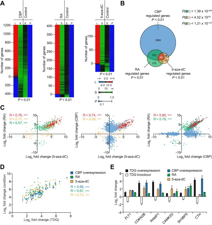
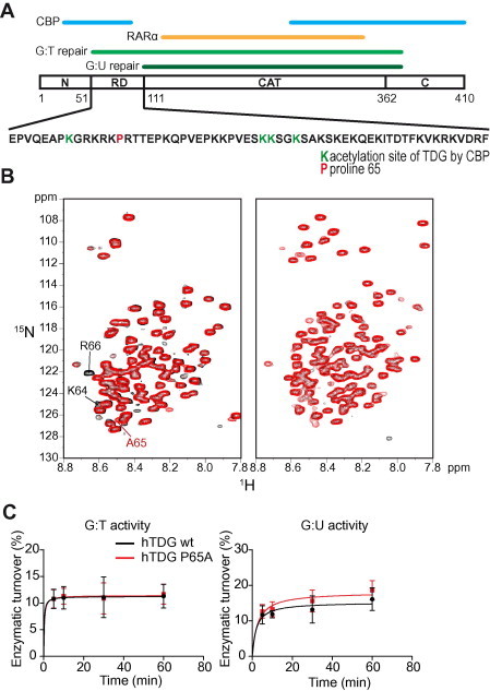
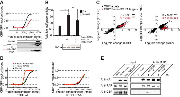
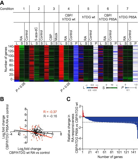
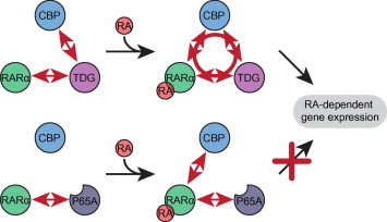
The thymine DNA glycosylase (TDG) is a multifunctional enzyme, which is essential for embryonic development. It mediates the base excision repair (BER) of G:T and G:U DNA mismatches arising from the deamination of 5-methyl cytosine (5-MeC) and cytosine, respectively. Recent studies have pointed at a role of TDG during the active demethylation of 5-MeC within CpG islands. TDG interacts with the histone acetylase CREB-binding protein (CBP) to activate CBP-dependent transcription. In addition, TDG also interacts with the retinoic acid receptor α (RARα), resulting in the activation of RARα target genes. Here we provide evidence for the existence of a functional ternary complex containing TDG, CBP and activated RARα. Using global transcriptome profiling, we uncover a coupling of de novo methylation-sensitive and RA-dependent transcription, which coincides with a significant subset of CBP target genes. The introduction of a point mutation in TDG, which neither affects overall protein structure nor BER activity, leads to a significant loss in ternary complex stability, resulting in the deregulation of RA targets involved in cellular networks associated with DNA replication, recombination and repair. We thus demonstrate for the first time a direct coupling of TDG's epigenomic and transcription regulatory function through ternary complexes with CBP and RARα.

Keywords: CREB-binding protein; Cytosine DNA methylation; Epigenomics; Retinoic acid receptor α; Thymine DNA glycosylase; Transcription regulation.


Figures

Figure 1
Figure 1
Comparison of CBP, 5-aza-dC and RA target genes by transcriptome profiling A. Heatmaps of a substraction profile of significantly (post hoc pFDR < 0.01) regulated genes after CBP overexpression (left), RA treatment (middle) and 5-aza-dC treatment (right) of HEK293 cells. Mock transfected, ethanol- or DMSO-treated cells were used as controls, respectively. The number of genes (L, column 1) that are positively (red shading) and negatively (blue shading) regulated is shown together with the average over three independent biological replicate signals for each condition (S, column 2) and control (S, column 3). The final column (column 4) represents the post hoc P values (P). B. Venn diagram for the overlap of genes regulated in a statistically significant manner in all three conditions studied in A. The P values (hypergeometric distribution) for chance occurrence of each overlap are indicated. Note that the overall overlap of 86 genes is almost entirely composed of genes which are derepressed by 5-aza-dC treatment and concomittantly activated by atRA treatment and CBP overexpression. C. Scatter plots of the overlaps of methylation and RA-regulated genes (left), methylation and CBP-regulated genes (middle), and RA- and CBP- regulated genes (right) reveals clear Pearson’s correlations (R). Genes with expression significantly affected under both conditions compared in each panel are indicated in red. Genes with expression significantly affected upon CBP overexpression only (blue), by RA treatment only (green) and by 5-aza-dC treatment only (brown) are also indicated in different colors. ∗∗∗< 0.001. D. Scatter-plot of the common subset from panel B in each condition (Y axis) compared to the overexpression of human TDG in HEK293 cells (X axis). E. Gene expression changes of an assigned set of the common subset from panel B in HEK293 cells overexpressing human TDG and in TDG knockout mouse embryonic fibroblasts . TDG, thymine DNA glycosylase; RA, retinoic acid; 5-aza-dC, 5-azacytidine; FLT1, fls-related tyrosine kinase 1; CDKN2B, cyclin-dependent kinase inhibitor 2B; RAMP1, receptor activity modifying protein 1; CAMK2D, calcium/calmodulin-dependent protein kinase II delta; SH3BP5, SH3-domain binding protein 5; CTH, cystathionase.
Figure 2
Figure 2
Structure and activity of wild type TDG and the mutant P65A A. Schematic structure of hTDG. The regions required for G:T and G:U mismatch recognition and processing (light and dark green, respectively), the N- and C-termini, the regulatory domain (RD), the catalytic (CAT) domain as well as the interfaces with CBP (blue) and RARα (brown) are indicated. The amino acid sequence of the RD is provided and the lysines that can be acetylated by CBP and the proline 65 that was mutated in this study are highlighted in green and red, respectively. B.1H–15N HSQC spectra of the N-terminus (amino acid residues 1–111) (left panel) and full length hTDG (right panel). Spectra for wild-type and the P65A mutant are shown in black and red, respectively. The resonances of the neighboring amino acids of P65 as well as the resonance of the introduced alanine are indicated. C. Glycosylase kinetics of human TDG wild-type and P65A mutant on G:T and G:U repair. DNA nicking assays were performed on a 25-mer dsDNA containing either a central G:T (left panel) or a G:U (right panel) mismatch. A 25-mer dsRNA containing a central canonical G:C pair was used as a control.
Figure 3
Figure 3
The TDG P65A mutation results in reduced TDG/CBP/RARα ternary complex stability A. A representative microscale thermophoresis (MST) experiment comparing HA-tagged hTDG wt to HA-tagged hTDG P65A. Ten successive 1:1 dilutions of immuno-purified HA-hTDG (wt or P65A mutant) in binding buffer were used for MST analyses with YFP-CBP. The amounts of HA-fusion protein in every second dilution were monitored by Western blot using anti-HA antibody. The protein concentration of the non-diluted HA-hTDG samples was measured by Bradford assay. B. Promoter activation assay using a minimal Gal4 promoter driving the expression of firefly luciferase. The reporter construct was co-transfected with a Gal4-RARα expression vector and, where indicated, with either wild-type or P65A mutant HA-hTDG. Luciferase activity was normalized by protein amounts. Comparable TDG protein expression in the different conditions was verified by Western blotting. ∗∗< 0.01 in a students t-test. C. Scatter plot of the genes with expression significantly (P < 0.01) regulated by CBP overexpression (X axis) upon overexpression of TDG (Y axis) wild-type (left panel) or TDG P65A (right panel). Pearson correlation coefficients for all CBP target genes were shown in black and those for the common subset with the 5-aza-dC and RA treatments from Figure 1D are shown in red. ∗∗∗< 0.001. D. MST experiments using increasing concentrations of purified HA-hTDG wild-type (left panel) or P65A mutant (right panel) in the presence of non-activated (green) or RA-activated (red) purified FLAG-tagged hRARα compared to HA-hTDG wt or P65A mutant alone (control). E. Immunoprecipitation of HA-TDG wt or P65A mutant from nuclear extracts of transiently transfected HEK293 cells in the presence or absence of RA using anti-HA agarose. Co-immnoprecipitated proteins were monitored for RAR and CBP using the appropriate antibodies. Mock transfected cells were used as control. Only input without RA treatment was included since addition of RA does not affect expression of the relevant proteins (data not shown).
Figure 4
Figure 4
TDG P65A influences the expression of genes sensitive to de novo DNA demethylation, RA and CBP A. Heatmap of significant (< 0.05) RA-responsive genes in non-transfected (Condition 1) and in CBP and wild-type hTDG overexpressing HEK293 cells (Condition 4). The number of genes (L) that are positively (red shading) or negatively (blue shading) regulated is shown together with the average over three independent biological replicate signals of each experimental condition and the corresponding control condition (S). Expression changes of these genes upon 5-aza-dC treatment (Condition 2), CBP overexpression (Condition 3) as well as RA treatment during simultaneous overexpression CBP and hTDG P65A (Condition 6) are displayed as heat maps. Cells overexpressing TDG wt (Condition 5) or P65A mutant (Condition 7) alone and subjected to RA treatment were used as control. Post-hoc P values are indicated as color code (P). B. Scatter plot of the genes shown in panel A. RA-mediated differential expression was compared between cells overexpressing CBP and hTDG wt (X axis) and cells overexpressing CBP and hTDG P65A mutant (Y axis). Genes with expression significantly (< 0.05) impacted in the hTDG P65A condition are highlighted in red. Pearson correlation coefficients for both non-significant (black) and significant subsets are indicated. C. Relative difference in RA-responsiveness of cells overexpressing CBP and hTDG P65A mutant in relative to cells overexpressing CBP and hTDG wt. The RA-responsiveness was normalized to the CBP/hTDG wt condition. Genes whose expression is significantly (P < 0.05) affected by RA treatment during CBP and hTDG P65A expression are highlighted in red, whereas genes with RA-dependent expression significantly (P < 0.05) affected during CBP and wild-type hTDG expression are highlighted in blue.
Figure 5
Figure 5
Model of ternary CBP:TDG:RAR complex action The interaction of CBP with RA-activated RARα cooperates with the interactions of TDG with CBP and RARα in order to stabilize a ternary complex composed of all three molecules, which affects the expression of RA-dependent genes (top). The P65A mutant of TDG is still able to interact with RARα, but fails to bind CBP, resulting in the inability to form a stable functional ternary complex (bottom).
None

Similar articles

Cited by

References

    1. Schär P., Fritsch O. DNA repair and the control of DNA methylation. Prog Drug Res. 2011;67:51–68. - PubMed
    1. Deem A.K., Li X., Tyler J.K. Epigenetic regulation of genomic integrity. Chromosoma. 2012;121:131–151. - PMC - PubMed
    1. Ehrlich M., Zhang X.Y., Inamdar N.M. Spontaneous deamination of cytosine and 5-methylcytosine residues in DNA and replacement of 5-methylcytosine residues with cytosine residues. Mutat Res. 1990;238:277–286. - PubMed
    1. Krokan H.E., Drabløs F., Slupphaug G. Uracil in DNA – occurrence, consequences and repair. Oncogene. 2001;21:8935–8948. - PubMed
    1. Ehrlich M. DNA hypomethylation in cancer cells. Epigenomics. 2009;1:239–259. - PMC - PubMed

Publication types

MeSH terms