

Inhibition of stearoyl-coA desaturase selectively eliminates tumorigenic Nanog-positive cells: improving the safety of iPS cell transplantation to myocardium
- PMID: 24394703
- PMCID: PMC3979912
- DOI: 10.4161/cc.27677
Inhibition of stearoyl-coA desaturase selectively eliminates tumorigenic Nanog-positive cells: improving the safety of iPS cell transplantation to myocardium
Abstract
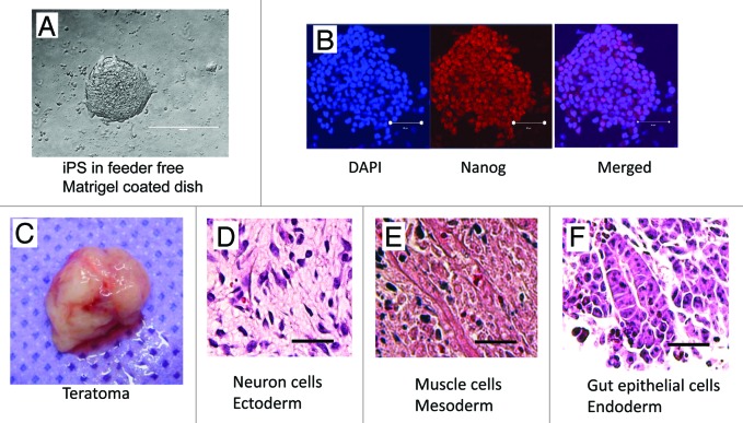
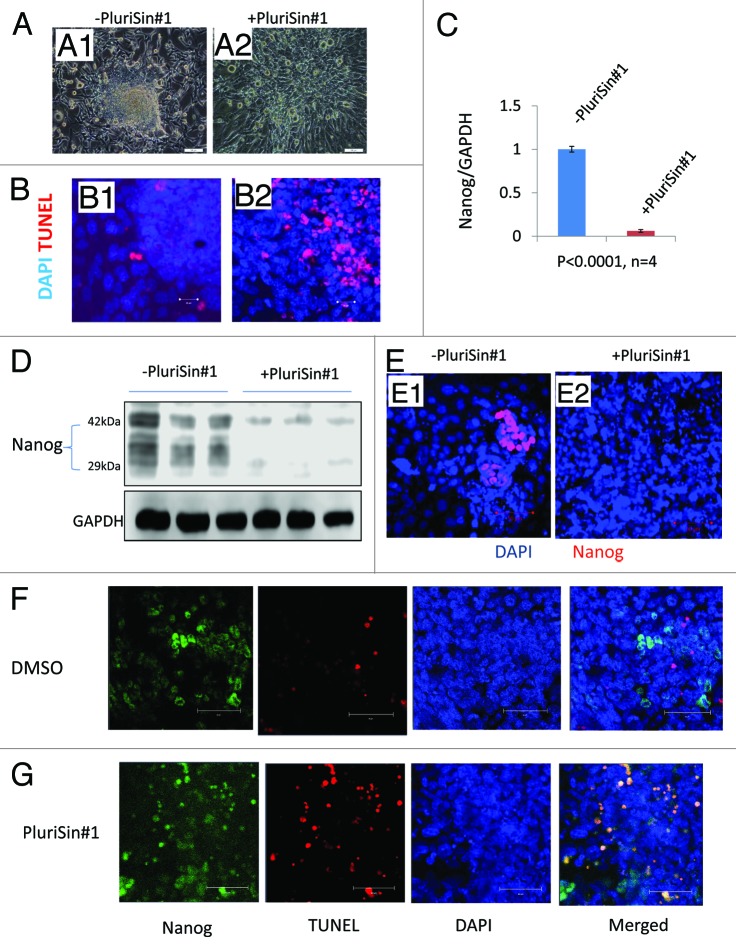
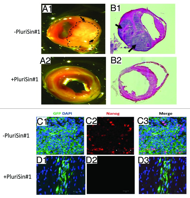
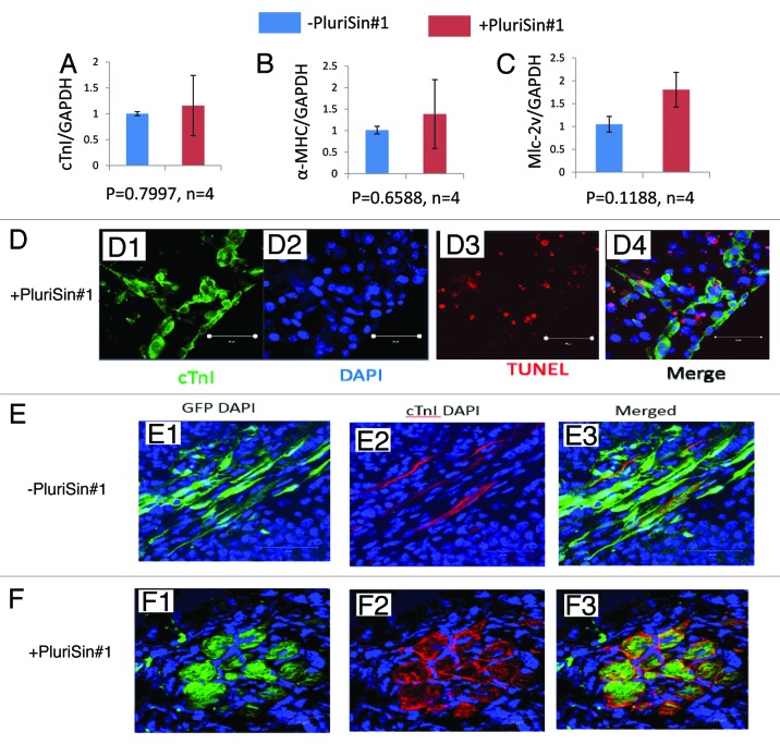
Induced pluripotent stem cells (iPS) can differentiate into cardiomyocytes (CM) and represent a promising form of cellular therapy for heart regeneration. However, residual undifferentiated iPS derivates (iPSD), which are not fully eliminated by cell differentiation or purification protocols, may form tumors after transplantation, thus compromising therapeutic application. Inhibition of stearoyl-coA desaturase (SCD) has recently been reported to eliminate undifferentiated human embryonic stem cells, which share many features with iPSD. Here, we tested the effects of PluriSin#1, a small-molecule inhibitor of SCD, on iPS-derived CM. We found that plurisin#1 treatment significantly decreased the mRNA and protein level of Nanog, a marker for both cell pluripotency and tumor progression; importantly, we provide evidence that PluriSin#1 treatment at 20 µM for 1 day significantly induces the apoptosis of Nanog-positive iPSD. In addition, PluriSin#1 treatment at 20 µM for 4 days diminished Nanog-positive stem cells in cultured iPSD while not increasing apoptosis of iPS-derived CM. To investigate whether PluriSin#1 treatment prevents tumorigenicity of iPSD after cell transplantation, we intramyocardially injected PluriSin#1- or DMSO-treated iPSD in a mouse model of myocardial infarction (MI). DMSO-treated iPSD readily formed Nanog-expressing tumors 2 weeks after injection, which was prevented by treatment with PluriSin#1. Moreover, treatment with PluriSin#1 did not change the expression of cTnI, α-MHC, or MLC-2v, markers of cardiac differentiation (P>0.05, n = 4). Importantly, pluriSin#1-treated iPS-derived CM exhibited the ability to engraft and survive in the infarcted myocardium. We conclude that inhibition of SCD holds the potential to enhance the safety of therapeutic application of iPS cells for heart regeneration.
Keywords: Nanog; PluriSin#1; iPS; myocardial infarction; tumorigenicity.
Figures

Comment in
-
Tumor-free iPS stem cells for heart cells.Cell Cycle. 2014;13(10):1519. doi: 10.4161/cc.28894. Epub 2014 Apr 22. Cell Cycle. 2014. PMID: 24755736 Free PMC article. No abstract available.
Similar articles
-
Tumor-free iPS stem cells for heart cells.Cell Cycle. 2014;13(10):1519. doi: 10.4161/cc.28894. Epub 2014 Apr 22. Cell Cycle. 2014. PMID: 24755736 Free PMC article. No abstract available.
-
Induced pluripotent stem (iPS) cells repair and regenerate infarcted myocardium.Mol Pharm. 2011 Oct 3;8(5):1573-81. doi: 10.1021/mp2001704. Epub 2011 May 24. Mol Pharm. 2011. PMID: 21542647 Free PMC article.
-
Neuregulin-1β induces mature ventricular cardiac differentiation from induced pluripotent stem cells contributing to cardiac tissue repair.Stem Cells Dev. 2015 Feb 15;24(4):484-96. doi: 10.1089/scd.2014.0211. Epub 2014 Nov 25. Stem Cells Dev. 2015. PMID: 25329043 Free PMC article.
-
[Regeneration of the central nervous system using iPS cell-technologies].Rinsho Shinkeigaku. 2009 Nov;49(11):825-6. doi: 10.5692/clinicalneurol.49.825. Rinsho Shinkeigaku. 2009. PMID: 20030221 Review. Japanese.
-
Generation of clinically relevant "induced pluripotent stem" (iPS) cells.J Stem Cells. 2011;6(3):109-27. J Stem Cells. 2011. PMID: 23264997 Review.
Cited by
-
A small molecule modulating monounsaturated fatty acids and Wnt signaling confers maintenance to induced pluripotent stem cells against endodermal differentiation.Stem Cell Res Ther. 2021 Oct 21;12(1):550. doi: 10.1186/s13287-021-02617-x. Stem Cell Res Ther. 2021. PMID: 34674740 Free PMC article.
-
FAK signaling suppression by OCT4-ITGA6 mediates the effectively removal of residual pluripotent stem cells and enhances application safety.Theranostics. 2025 Jun 12;15(14):7127-7153. doi: 10.7150/thno.111198. eCollection 2025. Theranostics. 2025. PMID: 40585989 Free PMC article.
-
Metabolic changes of human induced pluripotent stem cell-derived cardiomyocytes and teratomas after transplantation.iScience. 2024 Oct 23;27(11):111234. doi: 10.1016/j.isci.2024.111234. eCollection 2024 Nov 15. iScience. 2024. PMID: 39569381 Free PMC article.
-
Infrared fluorescent protein 1.4 genetic labeling tracks engrafted cardiac progenitor cells in mouse ischemic hearts.PLoS One. 2014 Oct 30;9(10):e107841. doi: 10.1371/journal.pone.0107841. eCollection 2014. PLoS One. 2014. PMID: 25357000 Free PMC article.
-
Report of the Key Opinion Leaders Meeting on Stem Cell-derived Beta Cells.Transplantation. 2018 Aug;102(8):1223-1229. doi: 10.1097/TP.0000000000002217. Transplantation. 2018. PMID: 29781950 Free PMC article. Review.
References
-
- Ohnuki M, Takahashi K, Yamanaka S. Generation and characterization of human induced pluripotent stem cells. Current protocols in stem cell biology 2009; Chapter 4:Unit 4A 2. - PubMed
Publication types
MeSH terms
Substances
Grants and funding
LinkOut - more resources
Full Text Sources
Other Literature Sources
Research Materials