. 2014 Mar 20;426(6):1285-95.
doi: 10.1016/j.jmb.2013.12.019. Epub 2014 Jan 4.

Domain interactions in adenovirus VAI RNA mediate high-affinity PKR binding

Affiliations

Domain interactions in adenovirus VAI RNA mediate high-affinity PKR binding

Katherine Launer-Felty et al. J Mol Biol. .

Abstract

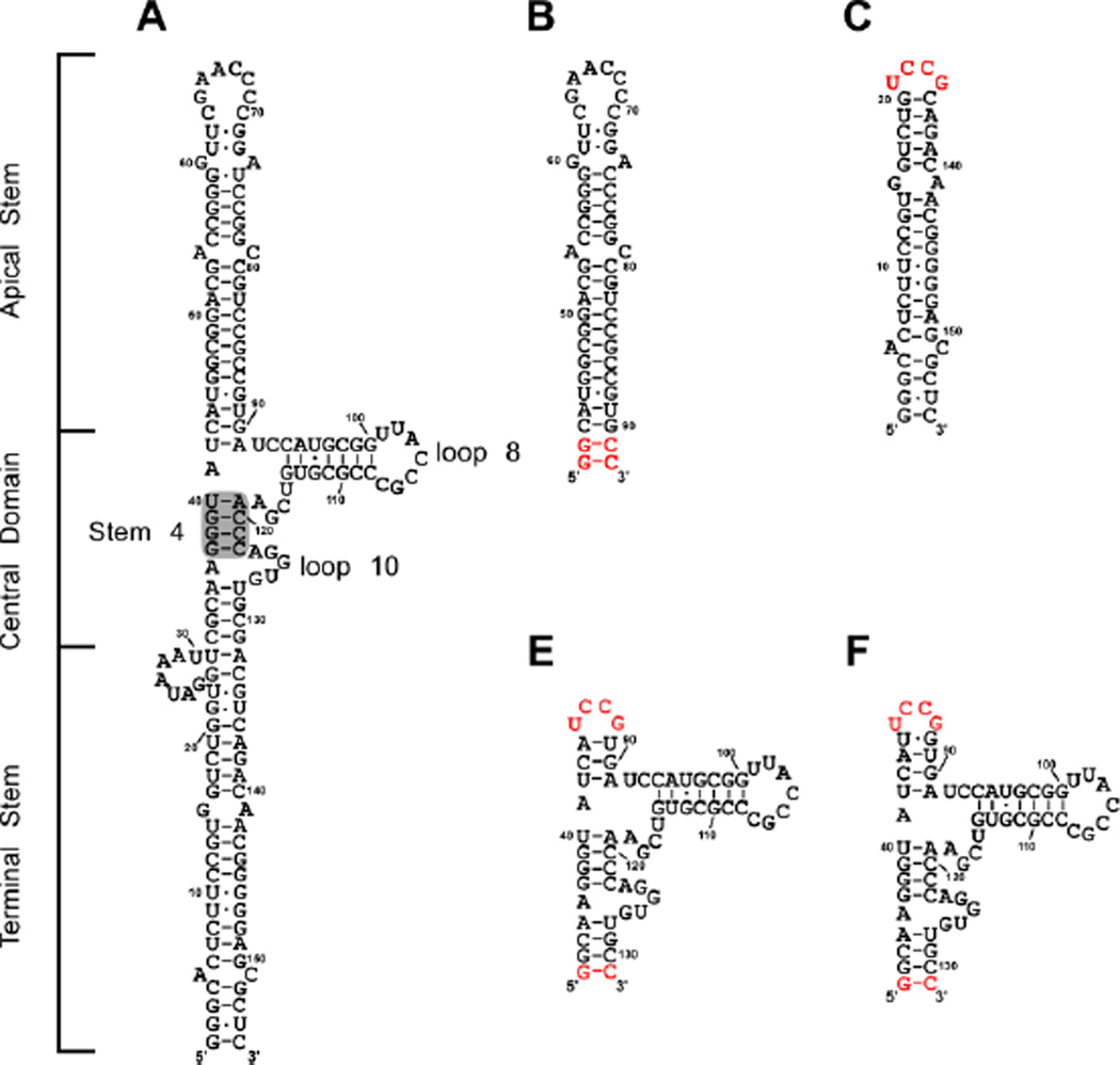
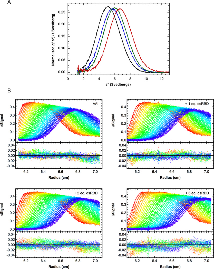
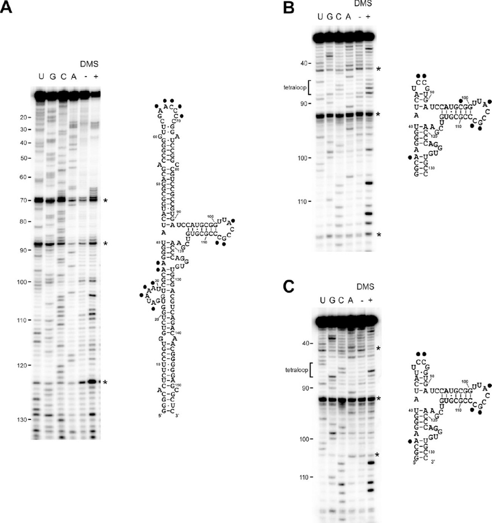
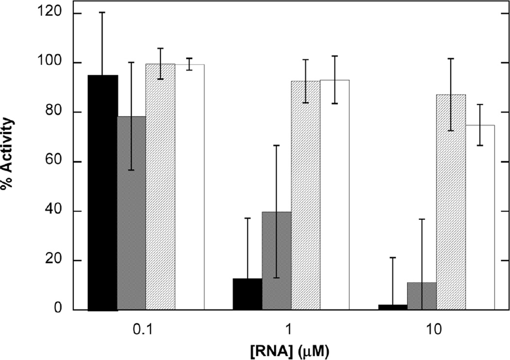
Protein kinase R (PKR) is a component of the innate immunity antiviral pathway. PKR is activated upon binding to double-stranded RNA (dsRNA) to undergo dimerization and autophosphorylation. Adenovirus-associated RNA I (VAI) is a short, non-coding transcript whose major function is to inhibit the activity of PKR. VAI contains three domains: an apical stem-loop, a highly structured central domain, and a terminal stem. Previous studies have localized PKR binding to the apical stem and to the central domain. However, the molecular mechanism for inhibition of PKR is not known. We have characterized the stoichiometry and affinity of PKR binding to VAI and several domain constructs using analytical ultracentrifugation and correlated VAI binding and PKR inhibition. Although PKR binding to simple dsRNAs is not regulated by divalent ion, analysis of the interaction of the isolated dsRNA binding domain with VAI reveals that the binding affinity is enhanced by divalent ion. Dissection of VAI into its constituent domains indicates that none of the isolated domains retains the PKR binding affinity or inhibitory potency of the full-length RNA. PKR is capable of binding the isolated terminal stem, but deletion of this domain from VAI does not affect PKR binding or inhibition. These results indicate that both the apical stem and the central domain are required to form a high-affinity PKR binding site. Our data support a model whereby VAI functions as a PKR inhibitor because it binds a monomer tightly but does not facilitate dimerization.

Keywords: DMS probing; analytical ultracentrifugation; innate immunity; protein kinase; protein–nucleic acid interactions.


Figures

Figure 1
Figure 1
Secondary structure of VAI and domain constructs. A) Secondary structure of full length VAI as proposed in reference 35. Stem 4, loop 8 and loop 10 are annotated as indicated in reference 30. Shaded region indicates conserved tetranucleotide pair. Secondary structures of B) apical stem (AS), C) terminal stem (TS), D) central domain-1 (CD1), E) central domain-2 (CD2). All domain constructs are numbered according to full length VAI. Bases in depicted in red denote those added for efficient in vitro transcription.
Figure 2
Figure 2
Sedimentation velocity analysis of dsRBD binding to VAI in the absence of Mg2+. A) Normalized g^(s*) distributions of 0.4 µM VAI (black), VAI + 1 eq. dsRBD (blue), VAI + 2 eq. dsRBD (green) and VAI + 6 eq. dsRBD (red). B) Global analysis of sedimentation velocity difference curves for dsRBD binding to VAI. The top panels show the data (points) and fit (solid lines) and the bottom panels show the residuals (points). The best fit parameters are shown in Table 1.
Figure 3
Figure 3
DMS structure probing of VAI (A), CD1 (B) and CD2 (C). Gels are shown on the left and secondary structures diagrams are shown on the right with modified bases indicated by a black dot. Each gel includes a sequencing ladder, a control lane without DMS (−) and a lane with DMS (+). Artifacts arising from reverse transcriptase pause sites are marked with an asterisk.
Figure 4
Figure 4
Sedimentation velocity analysis of PKR binding to VAI domain constructs. Normalized g^(s*) distributions for AS (A), TS (B), CD1 (C) and CD2 (D). For each construct the curves are labeled as follows: RNA alone (black), RNA + 1 eq. PKR (blue), RNA + 2 eq. PKR (green) and RNA + 6 eq. PKR (red).
Figure 5
Figure 5
Inhibition of PKR activation by VAI domain constructs. VAI (black), AS (gray), TS (diagonal lines), and CD2 (white). PKR was incubated with VAI domain constructs at the indicated concentration prior to addition of 10 µg/ml poly(rI:rC) and 0.4 mM ATP containing 4 µCi of γ-32P[ATP]. The percent inhibition was calculated relative to a sample containing no inhibitory RNA. The data represent the average of three experiments with error bars indicated.

References

    1. Unterholzner L, Bowie AG. The interplay between viruses and innate immune signaling: recent insights and therapeutic opportunities. Biochem. Pharmacol. 2008;75:589–602. - PubMed
    1. Weber F, Wagner V, Rasmussen SB, Hartmann R, Paludan SR. Double-stranded RNA is produced by positive-strand RNA viruses and DNA viruses but not in detectable amounts by negative-strand RNA viruses. Journal of Virology. 2006;80:5059–5064. - PMC - PubMed
    1. Langland JO, Cameron JM, Heck MC, Jancovich JK, Jacobs BL. Inhibition of PKR by RNA and DNA viruses. Virus Research. 2006;119:100–110. - PubMed
    1. Nanduri S, Carpick BW, Yang Y, Williams BR, Qin J. Structure of the double-stranded RNA binding domain of the protein kinase PKR reveals the molecular basis of its dsRNA-mediated activation. EMBO J. 1998;17:5458–5465. - PMC - PubMed
    1. Ryter JM, Schultz SC. Molecular basis of double-stranded RNA-protein interactions: structure of a dsRNA-binding domain complexed with dsRNA. EMBO J. 1998;17:7505–7513. - PMC - PubMed

Publication types

MeSH terms

LinkOut - more resources