. 2014 Jan 7:4:3573.
doi: 10.1038/srep03573.

Innexin gap junctions in nerve cells coordinate spontaneous contractile behavior in Hydra polyps

Affiliations

Innexin gap junctions in nerve cells coordinate spontaneous contractile behavior in Hydra polyps

Yasuharu Takaku et al. Sci Rep. .

Erratum in

  • Sci Rep. 2014;4:4009

Abstract

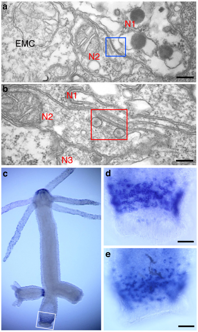
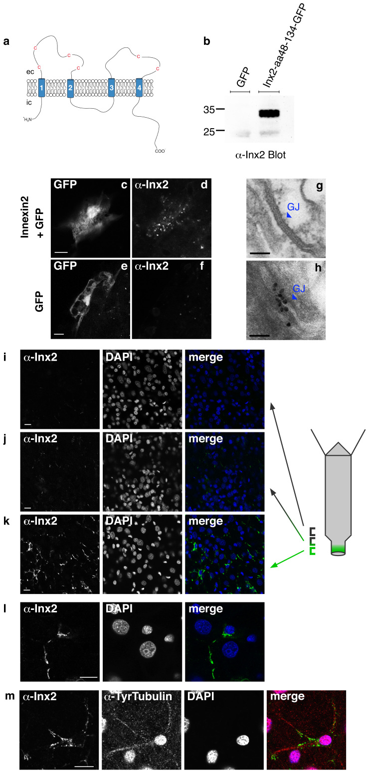
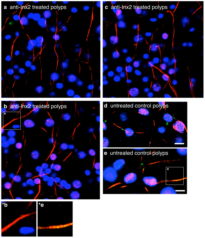
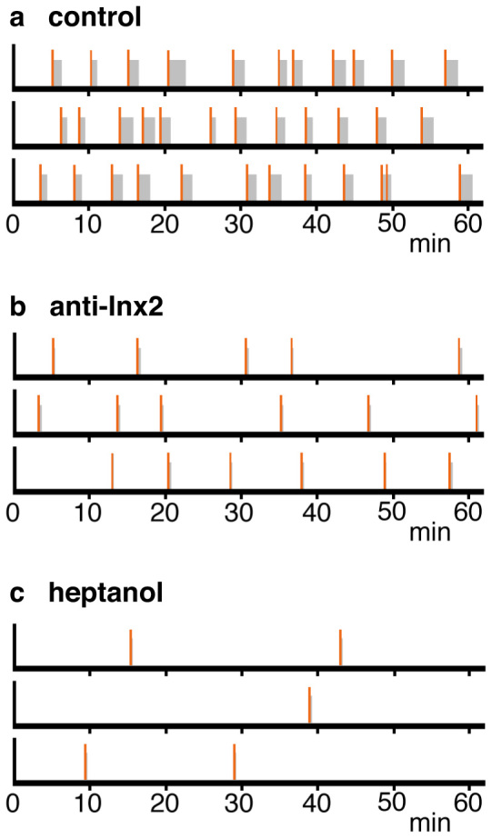
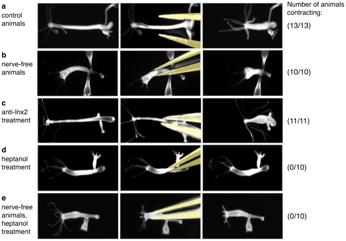
Nerve cells and spontaneous coordinated behavior first appeared near the base of animal evolution in the common ancestor of cnidarians and bilaterians. Experiments on the cnidarian Hydra have demonstrated that nerve cells are essential for this behavior, although nerve cells in Hydra are organized in a diffuse network and do not form ganglia. Here we show that the gap junction protein innexin-2 is expressed in a small group of nerve cells in the lower body column of Hydra and that an anti-innexin-2 antibody binds to gap junctions in the same region. Treatment of live animals with innexin-2 antibody eliminates gap junction staining and reduces spontaneous body column contractions. We conclude that a small subset of nerve cells, connected by gap junctions and capable of synchronous firing, act as a pacemaker to coordinate the contraction of the body column in the absence of ganglia.


Figures

Figure 1
Figure 1. Synapses between nerve cells and expression of innexin-2 in the peduncle of Hydra.
(a) and (b) TEM images of an electrical synapse (a) and a chemical synapse (b) between nerve cells in the peduncle. Squares outline the synapses. N1, N2, N3: nerve cells; EMC: epitheliomuscle cell. Scale bar, 200 nm. (c–e) Expression pattern of innexin-2 determined by whole mount in situ hybridization. The white box delineates the region of innexin-2 expression in the lower peduncle of the adult polyp. The fully developed bud on the left side also contains innexin-2 positive cells whereas the immature bud on the right side has not yet differentiated innexin-2 nerve cells. Staining in the tip of the hypostome appears to be background and is not associated with cellular structures. Enlarged peduncle regions of two additional polyps are shown in (d) and (e). Scale bar, 0.1 mm.
Figure 2
Figure 2. Immunofluorescent staining of innexin-2 in gap junctions in Hydra.
(a) Schematic drawing of the predicted structure of hydra innexin showing four transmembrane domains and conserved cysteine residues in the extracellular (ec) loops. N- and C-terminus are intracellular (ic). The first extracellular loop of innexin-2 (aa48–134) was used for antibody production. (b) Purified recombinant GFP-tagged innexin-2 (aa48–134) from E.coli was detected by the innexin-2 antibody at the appropriate size (ca. 35 kD) in immunoblot. The antibody did not detect GFP. (c–f) Ectopic expression of GFP (used as a transfection marker) and untagged innexin-2 in hydra epithelial cells transfected with the particle gun. To visualize innexin-2 expression, animals were fixed and stained with innexin-2 antibody 48 hours post-transfection. Transfected GFP-expressing epithelial cells displayed a punctate innexin-2 pattern in immunofluorescence when co-transfected with innexin-2 (d). Control animals transfected with GFP only have no detectable innexin-2 signal (f). Scale bar: 10 μm. (g) and (h) Immunogold staining of innexin-2 gap junction in peduncle tissue. (g) TEM image of a typical gap junction (enlarged from Fig. 1A); (h) immunogold staining of innexin-2 gap junction in peduncle tissue. Scale bar: 100 nm. (i–k) Immunostaining of whole animals with innexin-2 antibody revealed innexin-2 positive green spots primarily in the peduncle region (k); more apical areas in the peduncle showed decreased amounts of antibody staining (i and j); the gastric region contained no innexin-2 positive green spots. (l) High magnification image of an innexin-2 positive cell in the peduncle region. The innexin-2 positive green spots were often clustered as strings along nerve processes. Nuclei stained with DAPI. Projections of confocal images covering a depth of 2–3 μm. Scale bar: 10 μm. (m) Co-immunostaining of hydra with innexin-2 antibody and tyrosine-tubulin antibody (Sigma) showed that innexin-2 staining in nerve cells was localized along nerve cell processes. The anti-tyrosine-tubulin staining in nerve cell nuclei is regularly observed but appears to be an artifact (see also Figure 3). Nuclei stained with DAPI. Projections of confocal sections covering a depth of 2 μm. Scale bar: 10 μm.
Figure 3
Figure 3. Treatment of Hydra polyps with innexin-2 antibody eliminated innexin-2 stained gap junctions in peduncle tissue.
(a), (b) and (c) Confocal images of three anti-innexin-2 treated polyps fixed after 3 days and co-immunostained with innexin-2 antibody (green) and tyrosine-tubulin antibody (red). Nuclei are stained with DAPI (blue). (d) and (e) Confocal images of two DMSO treated control polyps. Note that innexin-2 spots (green) are yellow where directly overlapping with strong anti-tubulin (red) stained processes. (*b and *e) Enlargements of single nerve cell processes from b (anti-innexin-2 treated polyps) and e (untreated control polyps). Projections of confocal images covering a depth of 2 μm. Scale bar, 10 μm.
Figure 4
Figure 4. Role of innexin-2 gap junctions in contractile behavior.
(a) and (b) Schematic representation of contractile behavior over one hour interval based on time-lapse videos (see Supplemental movie 1 and 2). Data are shown for three individual polyps for each treatment. A single contraction is represented by a vertical bar (red); contraction bursts are represented by a vertical bar (red) followed by a grey bar showing the duration of the burst. Animals were treated with DMSO alone (control) or with DMSO and anti-innexin-2 antibody. (c) Contractile behavior of 3 individual polyps treated with 0.06% heptanol.
Figure 5
Figure 5. Contractile behavior induced by mechanical stimulation.
(a–e) Contraction of the body column was induced by pinching with forceps. Control hydra (a), nerve-free hydra (b), innexin-2 antibody treated hydra (c), heptanol treated normal hydra (d) and heptanol treated nerve-free hydra (e). The number of animals responding with body column contraction is shown on the right.

References

    1. Anderson P. A. V. Evolution of the first nervous systems. (Plenum Press, 1990).
    1. Galliot B. & Quiquand M. A two-step process in the emergence of neurogenesis. Eur. J. Neurosci. 34, 847–862 (2011). - PubMed
    1. Mackie G. O. The Elementary Nervous System Revisited. Am. Zool. 30, 907–920 (1990).
    1. Campbell R. D., Josephson R. K., Schwab W. E. & Rushforth N. B. Excitability of nerve-free hydra. Nature 262, 388–390 (1976). - PubMed
    1. Marder E. Electrical synapses: beyond speed and synchrony to computation. Curr. Biol. 8, R795–797 (1998). - PubMed

Publication types

LinkOut - more resources