

Ileal malignant hemangioendothelioma as a hypervascular lesion on computed tomography scan
- PMID: 24394857
- PMCID: PMC3907202
- DOI: 10.1016/j.ijscr.2013.09.019
Ileal malignant hemangioendothelioma as a hypervascular lesion on computed tomography scan
Abstract
Introduction: Malignant epithelioid hemangioendothelioma (EHE) is an uncommon and grave vascular tumor. EHE is frequently angiocentric and is associated with a medium sized vessel, especially a vein. No definite etiological associations have been ascribed to this tumor so far, except an association with oral contraceptives in EHE of liver.
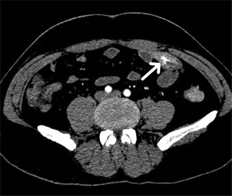
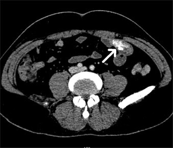
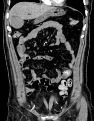
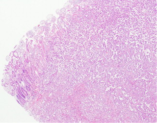
Presentation of case: A 47 year old man presented with the complaint of intermittent black stool over the past two weeks. Occasionally, he experienced pain in left lower abdomen. On Computed Tomography (CT), it showed hypervascular lesion in the ileum with persistent enhancement. An exploratory laparotomy was performed with short segmental resection and functional end-to-end anastomosis. It was diagnosed finally with the histopathological and immunohistochemical analysis as a malignant EHE.
Discussion: EHE is an uncommon endothelial tumor that most frequently arises in soft tissue, liver, lung and skeleton. It behaves biologically in between benign epithelioid hemangioma and the more aggressive epithelioid angiosarcoma. Although a standard systemic treatment for malignant EHE has not been fully established, complete surgical excision is strongly recommended if feasible.
Conclusion: EHE has a variable presentation and CT is helpful in identifying ileal EHE timely in the early stage, even when there is no obvious mass formation, however the diagnosis can be confirmed only after histopathological and immunohistochemical studies.
Keywords: Computed tomography (CT); Epithelioid hemangioendothelioma; Ileum; Immunohistochemical study; Prognosis.
Copyright © 2013 The Authors. Published by Elsevier Ltd.. All rights reserved.
Figures
Similar articles
-
Hepatic Epithelioid Hemangioendothelioma: Difficult Differential Diagnosis from Angiosarcoma.Case Rep Gastroenterol. 2020 Jan 29;14(1):56-62. doi: 10.1159/000505513. eCollection 2020 Jan-Apr. Case Rep Gastroenterol. 2020. PMID: 32110201 Free PMC article.
-
Malignant pleuropulmonary epithelioid hemangioendothelioma - unusual presentation of an aggressive angiogenic neoplasm.Pathol Res Pract. 2014 Sep;210(9):613-8. doi: 10.1016/j.prp.2014.04.011. Epub 2014 May 27. Pathol Res Pract. 2014. PMID: 24939148
-
Epithelioid hemangioendothelioma of the bone: A case report with findings of bone scintigraphy.Medicine (Baltimore). 2019 May;98(19):e15546. doi: 10.1097/MD.0000000000015546. Medicine (Baltimore). 2019. PMID: 31083212 Free PMC article.
-
Mediastinal Epithelioid Hemangioendothelioma Invading Superior Vena Cava: A Case Report and Review of Literature.Curr Med Imaging Rev. 2019;15(3):349-352. doi: 10.2174/1573405614666180124141817. Curr Med Imaging Rev. 2019. PMID: 31989887 Review.
-
[Research Progress of Pulmonary Epithelioid Hemangioendothelioma].Zhongguo Fei Ai Za Zhi. 2019 Jul 20;22(7):470-476. doi: 10.3779/j.issn.1009-3419.2019.07.10. Zhongguo Fei Ai Za Zhi. 2019. PMID: 31315787 Free PMC article. Review. Chinese.
Cited by
-
A rare case of intravascular epithelioid hemangioendothelioma of the cephalic vein treated with surgery and postoperative radiation therapy: a case report and review of the literature.J Med Case Rep. 2015 Apr 29;9:91. doi: 10.1186/s13256-015-0565-0. J Med Case Rep. 2015. PMID: 25924932 Free PMC article. Review.
References
-
- Yasuda S., Hashimoto T., Kanaizumi T., Kuwata H., Matsumoto I., Shiratori T. A case of hemangioendothelioma of the small intestine. Jpn J Surg. 1989;19(1):67–69. - PubMed
-
- Botsford T.W., Crowe P., Crocker D.W. Tumors of the small intestine. A review of experience with 115 cases including a report of a rare case of malignant hemangioendothelioma. Am J Surg. 1962;103:358–365. - PubMed
LinkOut - more resources
Full Text Sources
Other Literature Sources
Research Materials
Miscellaneous
