

Review
doi: 10.1002/dvdy.24110.
Epub 2014 Feb 27.
Rebirth of human embryology
Affiliations
- PMID: 24395627
- PMCID: PMC4310677
- DOI: 10.1002/dvdy.24110
Item in Clipboard
Review
Rebirth of human embryology
Dev Dyn.
2014 May.
No abstract available
Keywords: 3D reconstructions of systems; human embryology; labeled human embryo sections at every Stage.
Figures
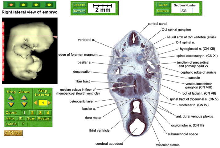
A typical page on the VHE-DREM website. The section image is from the Browse part of the database of a Stage 19 embryo. It is displayed with labels at the lowest magnification and includes the section number (#233) and scale bar. On the left is a side view of a computer generated reconstruction of the specimen’s surface with a section indicator showing the section level and plane that can be moved up or down to higher or lower section images. Below the surface reconstruction are buttons that change the magnification of the section image as well as access other parts of the database.
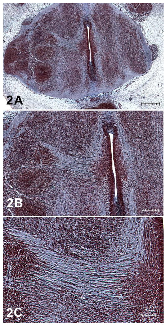
A Magnification level 2. Scale bar length equals 400 microns. B Magnification level 3. Scale bar length equals 200 microns. C Magnification level 4. Scale bar length equals 100 microns.
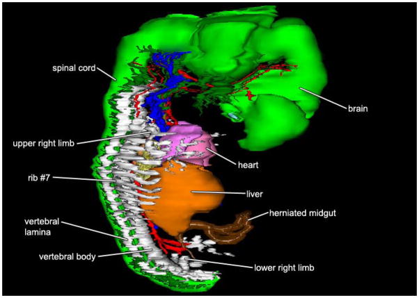
Right side view of a 3D reconstruction of the systems of the same embryo shown in Figure 1. Color code: brain, cranial nerves, and spinal cord: green; skeleton: white; heart: pink; arteries: red; veins: blue; lungs: yellow; liver: orange; midgut: brown.
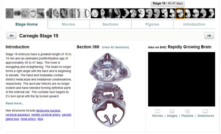
Example of a typical page from a VHE-DREM database as it appears on the EHD website. The unlabeled section image (#366) is from the same embryo shown in Figure 1. The text of the Introduction is displayed as an example on the left. Clicking on any one of the 27 specimens displayed along the top brings up the database for that specimen. Buttons displayed across the very top access movies, labeled serial section images, and figures of the stage displayed. At the bottom left is a list of the new structures that can be viewed in the appropriate section image by clicking on the hyperlink for that structure. An alphabetical index of all the labeled structures in all of the databases is available on this website.
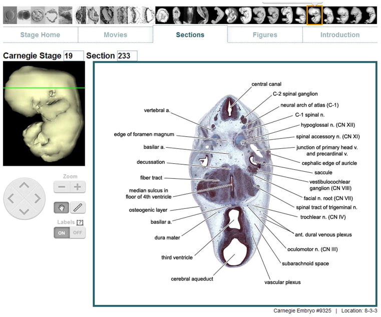
Example of a typical labeled section image (#366) from the same embryo shown in Figure 1 as it appears on the EHD website. At the top left is a side view of the reconstructed specimen on which the section level and plane are indicated. The navigation disk below it makes possible viewing the next higher or lower section image by activating the up or down arrowheads, respectively. Activating of the left or right arrowhead will access a similar section image from an earlier or later stage, respectively. To the right of the disk are buttons that raise or lower the magnification level of the section image, navigate the section area, take measurements, and turn on and off labels. Clicking the “?” displays definitions of all abbreviations used in the databases.
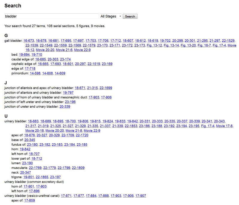
Example of a typical search results page on the EHD website showing a summary of all search results for the term “bladder” followed by each term and its related hyperlinks.
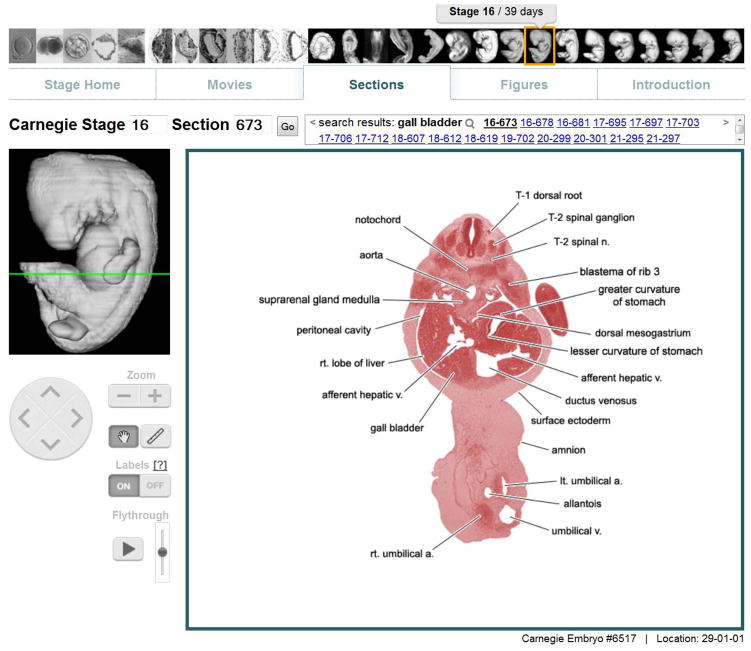
Example of EHD’s image viewer page after clicking the first hyperlink following “gall bladder” on the previous figure. Hyperlinks to all serial section images with a gall bladder label are listed in the search results box found just above the section image. This allows immediate access to every section image containing the search term. Clicking the magnifying glass will regenerate the search results page previously shown in Figure 6.
References
-
- Blechschmidt E. Die Pränatalin Organsysteme des Menschen. Stuttgart: Hipokrates Verlag; 1973.
-
- Blechschmidt E. The Beginning of Human Life. New York: Springer Verlag; 1977.
-
- Blechschmidt E, Gasser RF. Principles and Applications. Springfield, IL: Charles C. Thomas Publisher; 1978. Biokinetics and Biodynamics of Human Differentiation.
-
- Collins FS, Morgan M, Patrinos A. The Human Genome Project: Lessons from Large-Scale Biology. Science. 2003;300:286. - PubMed
-
- Dickey RP, Gasser RF. Variability in the growth rate of human embryos resulting from IVF and GIFT as determined by vaginal ultrasound. Anat Rec. 1993;236:47.
Publication types
MeSH terms
Grants and funding
LinkOut - more resources
Full Text Sources
Other Literature Sources
