

Enterocutaneous fistula 3 years after resection of an advanced gallbladder carcinoma
- PMID: 24395877
- PMCID: PMC3902497
- DOI: 10.1136/bcr-2013-202062
Enterocutaneous fistula 3 years after resection of an advanced gallbladder carcinoma
Abstract
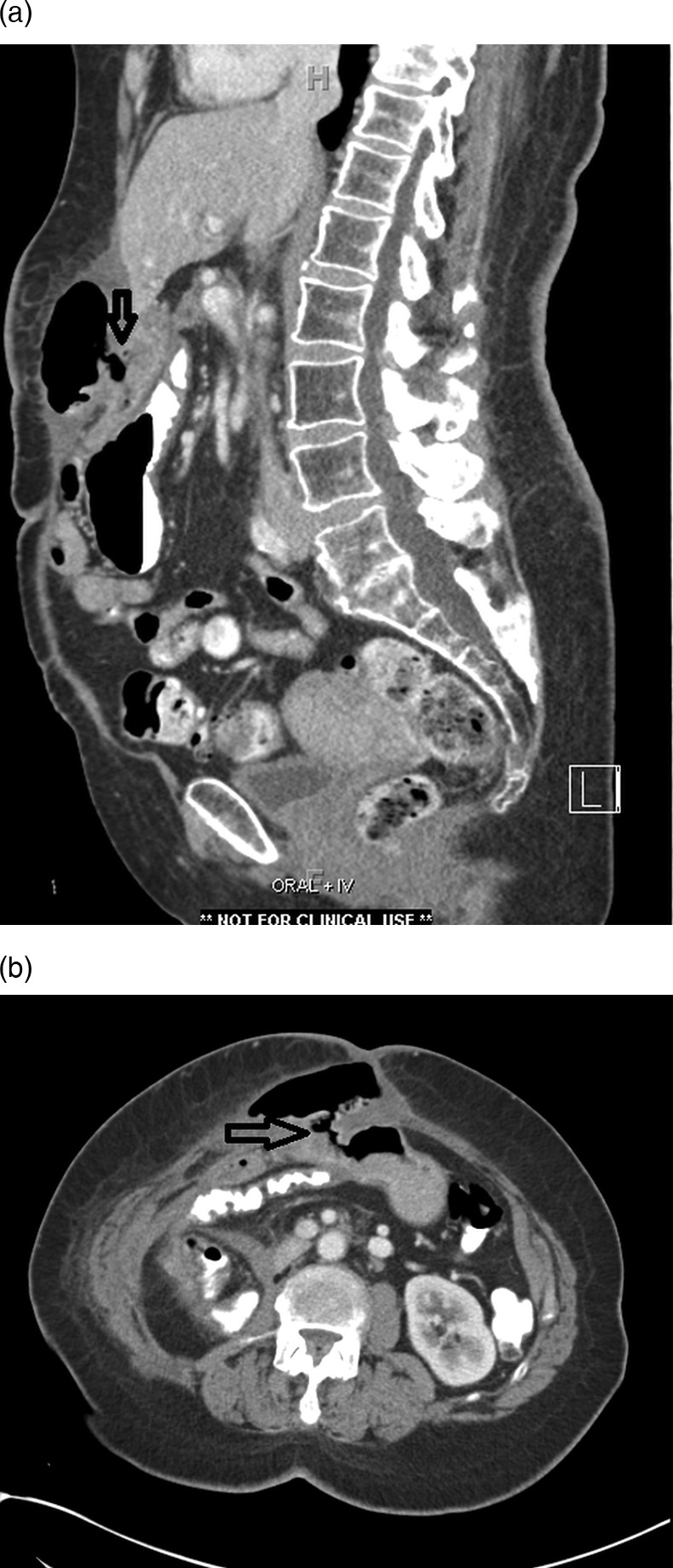
A 71-year-old woman presented to the emergency department with abdominal pain and fever. Her surgical history was significant for gallbladder adenocarcinoma for which she had undergone extensive resection 32 months previously. At that time she underwent cholecystectomy, wedge resection of the liver, pancreatoduodenectomy, right nephrectomy and right hemicolectomy for a locally advanced gallbladder adenocarcinoma. Examination revealed a tender, warm, upper midline abdominal wall mass. A CT scan with oral contrast revealed a fistulous tract extending from the gastrojejunostomy (GJ) into an abscess cavity in the adjacent anterior abdominal wall. She underwent open wound drainage with debridement, and was started on parenteral nutrition and intravenous antibiotics. The patient then underwent surgical repair excision of the fistula and refashioning of the GJ 1 month later. Histological examination of the specimen revealed well-healed suture lines, and no evidence of tumour recurrence.
Figures

References
-
- Berry SM, Fischer JE. Classification and pathophysiology of enterocutaneous fistulas. Surg Clin North Am 1996;76:1009–18 - PubMed
-
- Rubelowsky J, Machiedo GW. Reoperative versus conservative management for gastrointestinal fistulas. Surg Clin North Am 1991;71:147–57 - PubMed
-
- Garrido AB, Jr, Rossi M, Lima SE, Jr, et al. Early marginal ulcer following Roux-en-Y gastric bypass under proton pump inhibitor treatment: prospective multicentric study. Arq Gastroenterol 2010;47:130–4 - PubMed
-
- Csendes A, Burgos AM, Altuve J, et al. Incidence of marginal ulcer 1 month and 1 to 2 years after gastric bypass: a prospective consecutive endoscopic evaluation of 442 patients with morbid obesity. Obes Surg 2009;19:135–8 - PubMed
-
- Vasquez JC, Wayne Overby D, Farrell TM. Fewer gastrojejunostomy strictures and marginal ulcers with absorbable suture. Surg Endosc 2009;23:2011–15 - PubMed
Publication types
MeSH terms
LinkOut - more resources
Full Text Sources
Other Literature Sources
Medical