Review
. 2014 Jan;27(1):89-115.
doi: 10.1128/CMR.00070-13.

The endemic treponematoses

Affiliations
Review

The endemic treponematoses

Lorenzo Giacani et al. Clin Microbiol Rev. 2014 Jan.

Abstract

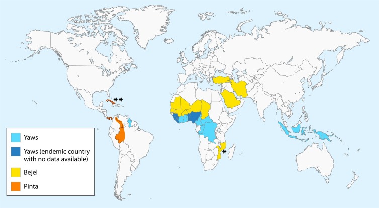
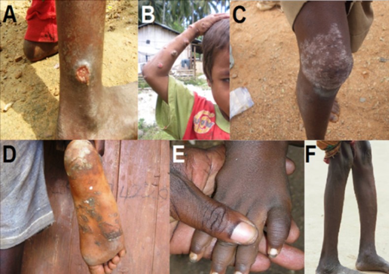
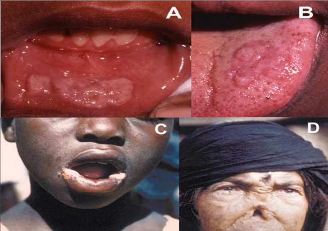
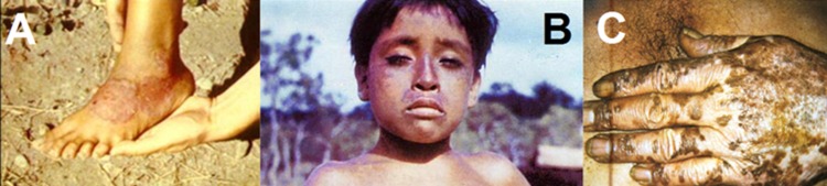
The agents of human treponematoses include four closely related members of the genus Treponema: three subspecies of Treponema pallidum plus Treponema carateum. T. pallidum subsp. pallidum causes venereal syphilis, while T. pallidum subsp. pertenue, T. pallidum subsp. endemicum, and T. carateum are the agents of the endemic treponematoses yaws, bejel (or endemic syphilis), and pinta, respectively. All human treponematoses share remarkable similarities in pathogenesis and clinical manifestations, consistent with the high genetic and antigenic relatedness of their etiological agents. Distinctive features have been identified in terms of age of acquisition, most common mode of transmission, and capacity for invasion of the central nervous system and fetus, although the accuracy of these purported differences is debated among investigators and no biological basis for these differences has been identified to date. In 2012, the World Health Organization (WHO) officially set a goal for yaws eradication by 2020. This challenging but potentially feasible endeavor is favored by the adoption of oral azithromycin for mass treatment and the currently focused distribution of yaws and endemic treponematoses and has revived global interest in these fascinating diseases and their causative agents.


Figures

FIG 1
FIG 1
Current geographical distribution of endemic treponematoses. The map was designed according to epidemiological data on yaws available at http://www.who.int/yaws/epidemiology/en/ and the gathered bibliography, including work by Harding (333), Bendel (334), and da Cruz-Ferreira and Sterneberg (335). *, based on a 2006 case report article describing two cases of bejel in children in Mozambique (267). **, based on an early pinta lesion identified in a female resident of Cuba visiting Austria (274). Officially, the last case of pinta in Cuba was reported in 1975.
FIG 2
FIG 2
Clinical manifestations of yaws. (A) Early (primary) yaws: infectious skin lesion known as “mother yaw” on the ankle. (B) Early (secondary) yaws: infectious ulcero-papillomatous lesions on the arm. (C) Early (secondary) yaws: scaly maculae of irregular shape on the knee. (D) Early (secondary) yaws: plantar hyperkeratosis with painful cracks and fissures. (E) Fusiform swelling of the fourth digit in a yaws patient with dactylitis. (F) Saber shin, caused by chronic osteitis. (Panels A, C, and D are courtesy of Cynthia Kwakye and the Ghana National Yaws Eradication Program, reproduced with permission. Panel B is courtesy of Christina Widaningrum-Mkes and the Ministry of Health of Indonesia, reproduced with permission. Panels E and F were originally taken by Laurent Ferradini and are reprinted from reference , published under a Creative Commons license.)
FIG 3
FIG 3
Clinical manifestations of bejel. (A) Early bejel: labial mucosal plaques. (B) Early bejel: lingual plaques. (C) Angular stomatitis of early bejel, which also may occur in yaws. (D) Late bejel rhinopharyngitis mutilans (gangosa), which also occurs in yaws. (Panels A and B are courtesy of Emmanuel Galoo, reprinted from reference with permission. Panel C is reprinted from reference with permission of the publisher. Panel D is reprinted from reference with permission.)
FIG 4
FIG 4
Clinical manifestations of pinta. (A) Early pinta: erythematous-squamous plaque. (B) Early pinta: hyperpigmented lesions of pinta. (C) Late pinta: depigmented lesions on hand. (Panels A and B are reprinted from reference with permission of the publisher. Panel C is reprinted from http://itg.content-e.eu/Generated/pubx/173/treponematoses/pinta.htm with permission from the Institute of Tropical Medicine-Antwerp).
None
None

References

    1. Miao RM, Fieldsteel AH. 1980. Genetic relationship between Treponema pallidum and Treponema pertenue, two noncultivable human pathogens. J. Bacteriol. 141:427–429 - PMC - PubMed
    1. Paster BJ. 2010. Phylum XV. Spirochaetes Garrity and Holt 2001, p 471–566 In Krieg NR, Ludwig W, Whitman WB, Hedlund BP, Paster BJ, Staley JT, Ward N, Brown D, Parte A. (ed), Bergey's manual of systematic bacteriology. Springer, New York, NY
    1. Magnuson HJ, Thomas EW, Olansky S, Kaplan BI, DeMello L, Cutler JC. 1956. Inoculation syphilis in human volunteers. Medicine 35:33–82. 10.1097/00005792-195602000-00002 - DOI - PubMed
    1. Magnuson HJ, Eagle H, Fleischman R. 1948. The minimal infectious inoculum of Spirochaeta pallida (Nichols Strain), and a consideration of its rate of multiplication in vivo. Am. J. Syphilis 32:1–18 - PubMed
    1. Turner TB, Hollander DH. 1957. Biology of the treponematoses. World Health Organization, Geneva, Switzerland - PubMed

Publication types

MeSH terms

LinkOut - more resources