

Therapeutic evaluation of microRNAs by molecular imaging
- PMID: 24396507
- PMCID: PMC3881098
- DOI: 10.7150/thno.4928
Therapeutic evaluation of microRNAs by molecular imaging
Abstract
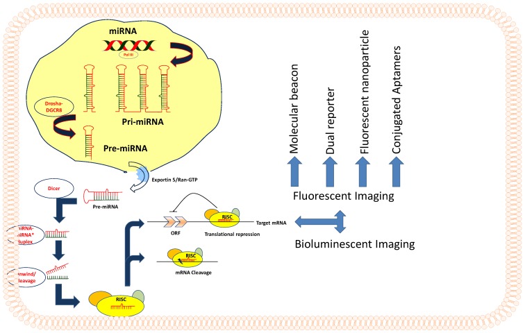
MicroRNAs (miRNAs) function as regulatory molecules of gene expression with multifaceted activities that exhibit direct or indirect oncogenic properties, which promote cell proliferation, differentiation, and the development of different types of cancers. Because of their extensive functional involvement in many cellular processes, under both normal and pathological conditions such as various cancers, this class of molecules holds particular interest for cancer research. MiRNAs possess the ability to act as tumor suppressors or oncogenes by regulating the expression of different apoptotic proteins, kinases, oncogenes, and other molecular mechanisms that can cause the onset of tumor development. In contrast to current cancer medicines, miRNA-based therapies function by subtle repression of gene expression on a large number of oncogenic factors, and therefore are anticipated to be highly efficacious. Given their unique mechanism of action, miRNAs are likely to yield a new class of targeted therapeutics for a variety of cancers. More than thousand miRNAs have been identified to date, and their molecular mechanisms and functions are well studied. Furthermore, they are established as compelling therapeutic targets in a variety of cellular complications. However, the notion of using them as therapeutic tool was proposed only recently, given that modern imaging methods are just beginning to be deployed for miRNA research. In this review, we present a summary of various molecular imaging methods, which are instrumental in revealing the therapeutic potential of miRNAs, especially in various cancers. Imaging methods have recently been developed for monitoring the expression levels of miRNAs and their target genes by fluorescence-, bioluminescence- and chemiluminescence-based imaging techniques. Mature miRNAs bind to the untranslated regions (UTRs) of the target mRNAs and regulate target genes expressions. This concept has been used for the development of fluorescent reporter-based imaging strategies to monitor the functional status of endogenous miRNAs, or the respective miRNAs transiently co-expressed in cells. Bioluminescence-based imaging strategies have been used to investigate various stages of miRNA processing and its involvement in different cellular processes. Similarly, chemiluminsecence methods were developed for in vitro miRNA imaging such as monitoring their therapeutic roles in various cancer cell lines.
Keywords: miRNAs; molecular Imaging.
Conflict of interest statement
Competing Interests: The authors have declared that no competing interest exists.
Figures
Similar articles
-
MicroRNAs as Therapeutic Targets and Colorectal Cancer Therapeutics.Adv Exp Med Biol. 2016;937:239-47. doi: 10.1007/978-3-319-42059-2_13. Adv Exp Med Biol. 2016. PMID: 27573904 Review.
-
microRNAs as oncogenes and tumor suppressors.Dev Biol. 2007 Feb 1;302(1):1-12. doi: 10.1016/j.ydbio.2006.08.028. Epub 2006 Aug 16. Dev Biol. 2007. PMID: 16989803 Review.
-
Developing therapeutic microRNAs for cancer.Gene Ther. 2011 Dec;18(12):1121-6. doi: 10.1038/gt.2011.79. Epub 2011 Jun 2. Gene Ther. 2011. PMID: 21633392 Free PMC article. Review.
-
MicroRNAs: non coding pleiotropic factors in development, cancer prevention and treatment.Microrna. 2013;2(2):81. doi: 10.2174/2211536611302020001. Microrna. 2013. PMID: 25070777
-
Molecular pathways: microRNAs as cancer therapeutics.Clin Cancer Res. 2012 Aug 15;18(16):4234-9. doi: 10.1158/1078-0432.CCR-11-2010. Epub 2012 Jun 18. Clin Cancer Res. 2012. PMID: 22711704 Free PMC article. Review.
Cited by
-
Genetically Encoded Reporter Genes for MicroRNA Imaging in Living Cells and Animals.Mol Ther Nucleic Acids. 2020 Sep 4;21:555-567. doi: 10.1016/j.omtn.2020.06.021. Epub 2020 Jun 27. Mol Ther Nucleic Acids. 2020. PMID: 32721876 Free PMC article. Review.
-
Bioluminescence Imaging for Monitoring miR-200c Expression in Breast Cancer Cells and its Effects on Epithelial-Mesenchymal Transition Progress in Living Animals.Mol Imaging Biol. 2018 Oct;20(5):761-770. doi: 10.1007/s11307-018-1180-4. Mol Imaging Biol. 2018. PMID: 29532351
-
Positive radionuclide imaging of miRNA expression using RILES and the human sodium iodide symporter as reporter gene is feasible and supports a protective role of miRNA-23a in response to muscular atrophy.PLoS One. 2017 May 11;12(5):e0177492. doi: 10.1371/journal.pone.0177492. eCollection 2017. PLoS One. 2017. PMID: 28493972 Free PMC article.
-
MicroRNAs in Schizophrenia: Implications for Synaptic Plasticity and Dopamine-Glutamate Interaction at the Postsynaptic Density. New Avenues for Antipsychotic Treatment Under a Theranostic Perspective.Mol Neurobiol. 2015 Dec;52(3):1771-1790. doi: 10.1007/s12035-014-8962-8. Epub 2014 Nov 14. Mol Neurobiol. 2015. PMID: 25394379 Review.
-
Biomarker-based MicroRNA Therapeutic Strategies for Hepatocellular Carcinoma.J Clin Transl Hepatol. 2014 Dec;2(4):253-8. doi: 10.14218/JCTH.2014.00020. Epub 2014 Dec 15. J Clin Transl Hepatol. 2014. PMID: 26355266 Free PMC article. Review.
References
-
- Ambros V. MicroRNA pathways in flies and worms: growth, death, fat, stress, and timing. Cell. 2003;113(6):673–676. - PubMed
-
- Bartel DP, Chen CZ. Micromanagers of gene expression: the potentially widespread influence of metazoan microRNAs. Nat Rev Genet. 2004;5(5):396–400. - PubMed
-
- Lee Y, Ahn C, Han J, Choi H, Kim J, Yim J, Lee J, Provost P, Radmark O, Kim S. et al. The nuclear RNase III Drosha initiates microRNA processing. Nature. 2003;425(6956):415–419. - PubMed
Publication types
MeSH terms
Substances
Grants and funding
LinkOut - more resources
Full Text Sources
Other Literature Sources
