

Gold nanoshelled liquid perfluorocarbon magnetic nanocapsules: a nanotheranostic platform for bimodal ultrasound/magnetic resonance imaging guided photothermal tumor ablation
- PMID: 24396512
- PMCID: PMC3881224
- DOI: 10.7150/thno.7275
Gold nanoshelled liquid perfluorocarbon magnetic nanocapsules: a nanotheranostic platform for bimodal ultrasound/magnetic resonance imaging guided photothermal tumor ablation
Abstract
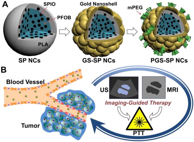
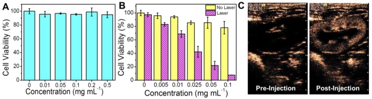
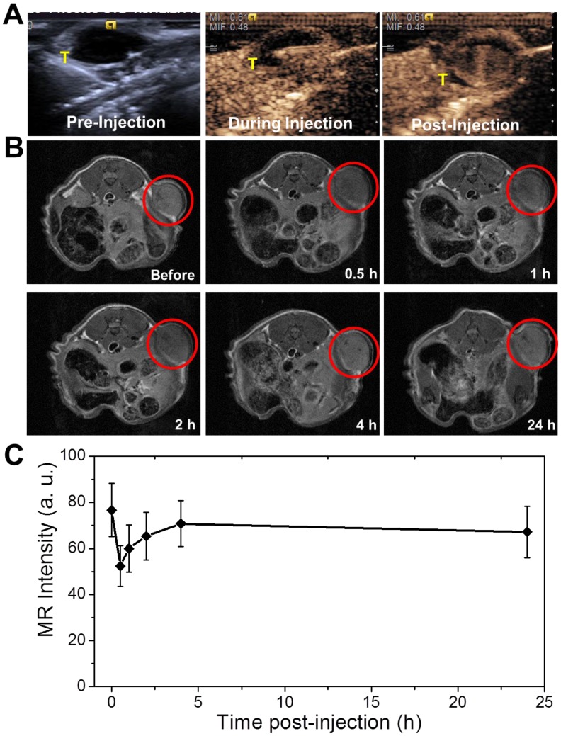
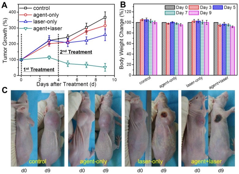
Imaging guided ablation therapy has been applied in both biomedical research and clinical trials and turned out to be one of the most promising approaches for cancer treatment. Herein, the multifunctional nanocapsules were fabricated through loading perfluorooctylbromide (PFOB) and superparamagnetic iron oxide nanoparticles (SPIOs) into poly(lactic acid) (PLA) nanocapsules (NCs), followed by the formation of PEGylated gold nanoshell on the surface. The resulting multi-component NCs were proved to be able to act as nanotheranostic agent to achieve successful bimodal ultrasound (US)/magnetic resonance imaging (MRI) guided photothermal ablation in human tumor xenograft models non-invasively. Such a single theranostic agent with the combination of real-time US and high-resolution MR imaging would be of great value to offer more comprehensive diagnostic information and dynamics of disease progression for the accurate location of therapeutic focusing spot in the targeted tumor tissue, showing great potential as an effective nanoplatform for contrast imaging guided photothermal therapy.
Keywords: Bimodal imaging; Gold nanoshell; Liquid perfluorocarbon nanocapules; Photothermal therapy.; Superparamagnetic iron oxide nanoparticles.
Conflict of interest statement
Competing Interests: The authors have declared that no competing interest exists.
Figures
Similar articles
-
Targeted gold nanoshelled hybrid nanocapsules encapsulating doxorubicin for bimodal imaging and near-infrared triggered synergistic therapy of Her2-positve breast cancer.J Biomater Appl. 2020 Sep;35(3):430-445. doi: 10.1177/0885328220929616. Epub 2020 Jun 9. J Biomater Appl. 2020. PMID: 32515640
-
Gold nanoshelled liquid perfluorocarbon nanocapsules for combined dual modal ultrasound/CT imaging and photothermal therapy of cancer.Small. 2014 Mar 26;10(6):1220-7. doi: 10.1002/smll.201302252. Epub 2014 Feb 5. Small. 2014. PMID: 24500926
-
Graphene Oxide and Gadolinium-Chelate Functionalized Poly(lactic acid) Nanocapsules Encapsulating Perfluorooctylbromide for Ultrasound/Magnetic Resonance Bimodal Imaging Guided Photothermal Ablation of Cancer.J Nanosci Nanotechnol. 2016 Mar;16(3):2201-9. doi: 10.1166/jnn.2016.10950. J Nanosci Nanotechnol. 2016. PMID: 27455619
-
A light-controllable specific drug delivery nanoplatform for targeted bimodal imaging-guided photothermal/chemo synergistic cancer therapy.Acta Biomater. 2018 Oct 15;80:308-326. doi: 10.1016/j.actbio.2018.09.024. Epub 2018 Sep 19. Acta Biomater. 2018. PMID: 30240955
-
Phase-Shifted PFH@PLGA/Fe3O4 Nanocapsules for MRI/US Imaging and Photothermal Therapy with near-Infrared Irradiation.ACS Appl Mater Interfaces. 2015 Jul 8;7(26):14231-42. doi: 10.1021/acsami.5b01873. Epub 2015 Jun 23. ACS Appl Mater Interfaces. 2015. PMID: 26067333
Cited by
-
Perfluorooctylbromide nanoparticles for ultrasound imaging and drug delivery.Int J Nanomedicine. 2018 May 25;13:3053-3067. doi: 10.2147/IJN.S164905. eCollection 2018. Int J Nanomedicine. 2018. PMID: 29872293 Free PMC article. Review.
-
"One-Pot" Fabrication of Highly Versatile and Biocompatible Poly(vinyl alcohol)-porphyrin-based Nanotheranostics.Theranostics. 2017 Sep 5;7(16):3901-3914. doi: 10.7150/thno.20190. eCollection 2017. Theranostics. 2017. PMID: 29109786 Free PMC article.
-
Ultrasound imaging beyond the vasculature with new generation contrast agents.Wiley Interdiscip Rev Nanomed Nanobiotechnol. 2015 Jul-Aug;7(4):593-608. doi: 10.1002/wnan.1326. Epub 2015 Jan 8. Wiley Interdiscip Rev Nanomed Nanobiotechnol. 2015. PMID: 25580914 Free PMC article. Review.
-
Superparamagnetic core/shell GoldMag nanoparticles: size-, concentration- and time-dependent cellular nanotoxicity on human umbilical vein endothelial cells and the suitable conditions for magnetic resonance imaging.J Nanobiotechnology. 2015 Mar 25;13:24. doi: 10.1186/s12951-015-0080-x. J Nanobiotechnology. 2015. PMID: 25890315 Free PMC article.
-
Laser activatable perfluorocarbon bubbles for imaging and therapy through enhanced absorption from coupled silica coated gold nanoparticles.RSC Adv. 2021 Jan 29;11(9):4906-4920. doi: 10.1039/d0ra08009h. eCollection 2021 Jan 25. RSC Adv. 2021. PMID: 35424456 Free PMC article.
References
-
- Huang X, El-Sayed IH, Qian W. et al. Cancer cell imaging and photothermal therapy in the near-infrared region by using gold nanorods. J Am Chem Soc. 2006;128:2115–20. - PubMed
-
- O'Neal DP, Hirsch LR, Halas NJ. et al. Photo-thermal tumor ablation in mice using near infrared-absorbing nanoparticles. Cancer Lett. 2004;209:171–6. - PubMed
-
- Loo C, Lowery A, Halas N. et al. Immunotargeted nanoshells for integrated cancer imaging and therapy. Nano Lett. 2005;5:709–11. - PubMed
Publication types
MeSH terms
Substances
LinkOut - more resources
Full Text Sources
Other Literature Sources
