. 2013 Jan 7;7(1):11802.
doi: 10.1063/1.4774311. eCollection 2013.

Size-based hydrodynamic rare tumor cell separation in curved microfluidic channels

Affiliations

Size-based hydrodynamic rare tumor cell separation in curved microfluidic channels

Jiashu Sun et al. Biomicrofluidics. .

Abstract

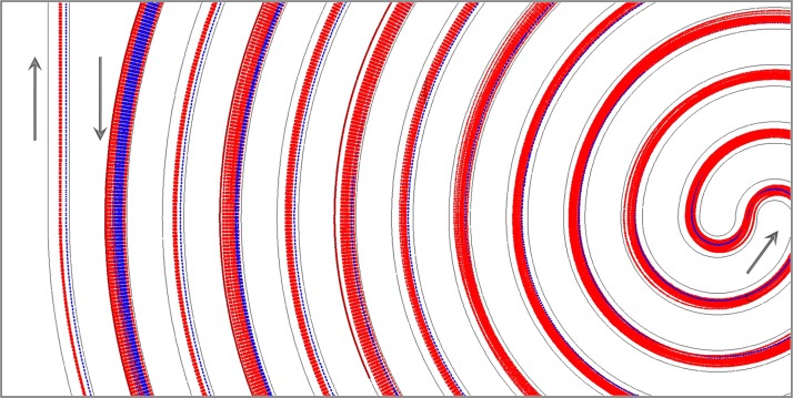
In this work, we propose a rapid and continuous rare tumor cell separation based on hydrodynamic effects in a label-free manner. The competition between the inertial lift force and Dean drag force inside a double spiral microchannel results in the size-based cell separation of large tumor cells and small blood cells. The mechanism of hydrodynamic separation in curved microchannel was investigated by a numerical model. Experiments with binary mixture of 5- and 15-μm-diameter polystyrene particles using the double spiral channel showed a separation purity of more than 95% at the flow rate above 30 ml/h. High throughput (2.5 × 10(8) cells/min) and efficient cell separation (more than 90%) of spiked HeLa cells and 20 × diluted blood cells was also achieved by the double spiral channel.


Figures

Figure 1
Figure 1
(a) Schematics of the microfluidic cell sorter containing 6-loop double spiral microchannel for particle/cell separation. (b) Picture of the assembled cell sorter.
Figure 2
Figure 2
Comparison of the Dean vortices and their magnitude at (a) 10 ml/h, and (b) 60 ml/h at the S-shape junction. The small arrows represent the velocity vector projected onto the cross-section and color contours represent the magnitude of the Dean flow velocity. The big red arrow indicates the flow direction.
Figure 3
Figure 3
Simulating prediction of trajectories of two types of particles near the inner/middle/outer outlet of the double spiral microchannel. (a) Flow rate 10 ml/h; (b) flow rate 20 ml/h; (c) flow rate 30 ml/h; (d) flow rate 40 ml/h; (e) flow rate 50 ml/h; (f) flow rate 60 ml/h. The red and blue dashed lines denote 5 μm particles and 15 μm particles, respectively.
Figure 4
Figure 4
Numerical prediction of the trajectories of 5 - and 15 -μm particles along the double spiral channel at the flow rate of 60 ml/h. The red and blue dashed lines denote 5 μm particles and 15 μm particles, respectively. The arrow represents the flow direction.
Figure 5
Figure 5
(a)–(f) Superimposed fluorescent images at the center of the double spiral channel for 5 μm (red line) and 15 μm (green line) particles at six different flow rates ranging from 10 to 60 ml/h. The arrow indicates the flow direction. (g) Line scans across the 300 μm wide channel of each composite image taken at the center to illustrate the distribution and position of 5 μm and 15 μm particles. (a′)–(f′) Superimposed fluorescent images at the outlet of the double spiral channel for 5 μm (red line) and 15 μm (green line) particles at the same flow rates. (g′) Line scans across the 300 μm wide channel of each composite image taken at the outlet. (a″)–(f″) Superimposed fluorescent images at the outlet of the single spiral channel. (g″) Line scans across the outlet of the single spiral channel.
Figure 6
Figure 6
(a) The optical microscope image indicating the trajectories of blood cells, (b) the fluorescent microscope image of HeLa cells, and (c) the superimposed image at the outlet of the double spiral channel at 40 ml/h. HeLa cells are stained with green and 20 × diluted blood cells are unstained. (a′)–(c′) The optical, fluorescent and superimposed microscope images taken at the outlet of the double spiral channel at 60 ml/h. (d) The composite image of cells collected at the middle outlet of the double spiral at 60 ml/h. (e) The composite image of cells collected at the inner outlet at 60 ml/h.

References

    1. Krivacic R. T., Ladanyi A., Curry D. N., Hsieh H. B., Kuhn P., Bergsrud D. E., Kepros J. F., Barbera T., Ho M. Y., Chen L. B., Lerner R. A., and Bruce R. H., Proc. Natl. Acad. Sci. U.S.A. 101, 10501 (2004).10.1073/pnas.0404036101 - DOI - PMC - PubMed
    1. Vincent M. E., Liu W. S., Haney E. B., and Ismagilov R. F., Chem. Soc. Rev. 39, 974 (2010).10.1039/b917851a - DOI - PMC - PubMed
    1. Hayes D. F., Miller M. C., Cristofanilli M., Ellis M. J., Stopek A., Allard W. J., Matera J., Doyle G. V., Terstappen L. W. W. M., and Budd G. T., Breast Cancer Res. Treat. 88, S225 (2004).
    1. Bell D. W. and Haber D. A., Clin. Cancer Res. 12, 3875 (2006).10.1158/1078-0432.CCR-06-0670 - DOI - PubMed
    1. Mostert B., Sleijfer S., Foekens J. A., and Gratama J. W., Cancer Treat. Rev. 35, 463 (2009).10.1016/j.ctrv.2009.03.004 - DOI - PubMed