Review
. 2013 Dec 14:2:44-51.
doi: 10.1016/j.redox.2013.12.003.

Redox regulation of the proteasome via S-glutathionylation

Affiliations
Review

Redox regulation of the proteasome via S-glutathionylation

Marilene Demasi et al. Redox Biol. .

Abstract

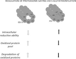
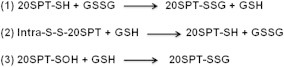
The proteasome is a multimeric and multicatalytic intracellular protease responsible for the degradation of proteins involved in cell cycle control, various signaling processes, antigen presentation, and control of protein synthesis. The central catalytic complex of the proteasome is called the 20S core particle. The majority of these are flanked on one or both sides by regulatory units. Most common among these units is the 19S regulatory unit. When coupled to the 19S unit, the complex is termed the asymmetric or symmetric 26S proteasome depending on whether one or both sides are coupled to the 19S unit, respectively. The 26S proteasome recognizes poly-ubiquitinylated substrates targeted for proteolysis. Targeted proteins interact with the 19S unit where they are deubiquitinylated, unfolded, and translocated to the 20S catalytic chamber for degradation. The 26S proteasome is responsible for the degradation of major proteins involved in the regulation of the cellular cycle, antigen presentation and control of protein synthesis. Alternatively, the proteasome is also active when dissociated from regulatory units. This free pool of 20S proteasome is described in yeast to mammalian cells. The free 20S proteasome degrades proteins by a process independent of poly-ubiquitinylation and ATP consumption. Oxidatively modified proteins and other substrates are degraded in this manner. The 20S proteasome comprises two central heptamers (β-rings) where the catalytic sites are located and two external heptamers (α-rings) that are responsible for proteasomal gating. Because the 20S proteasome lacks regulatory units, it is unclear what mechanisms regulate the gating of α-rings between open and closed forms. In the present review, we discuss 20S proteasomal gating modulation through a redox mechanism, namely, S-glutathionylation of cysteine residues located in the α-rings, and the consequence of this post-translational modification on 20S proteasomal function.

Keywords: 20SPT, 20S proteasome core particle; 26SPT, 26S proteasome; GSH, reduced glutathione; GSSG, oxidized glutathione; Oxidized proteins; Proteasomal gating; Proteasome; ROS, reactive oxygen species; Redox regulation; S-glutathionylation; SAXS, small angle X-rays scattering; TEM, transmission electron microscopy.


Figures

None
Graphical abstract
Fig. 1
Fig. 1
Major mechanisms of proteinS-glutathionylation. Route 1 describes the classical mechanism of protein S-glutathionylation. This mechanism causes modification through thiol–disulfide exchange. Formation of adduct may be triggered by an increased GSSG pool mediated by intensified ROS formation or the oxidized glutathione species GSOH and GS(O)SG. Route 2 describes mechanisms based on protein sulfenic acid formation through reaction with peroxides or peroxynitrite followed by reaction with GSH. Other physiological oxidants, e.g., hypochlorous acid or chloramine derivatives can also oxidize thiol groups to the sulfenic form . Route 3 describes mechanisms based on nitrosylated protein or gluathionyl derivatives that are formed through reaction with intermediates of NO radical metabolism.
Fig. 2
Fig. 2
Modeling of the 20SPT redox forms according to SAXS analyzes. (A) Top (left) and front (right) views of the 20SPT purified from yeast cells grown under conditions that promote fermentation; (B) same preparations after treatment with DTT. Internal and external diameters of the catalytic chamber (upper models) and the length of the 20SPT (botton left models) in both redox conditions were obtained through SAXS measurements . Modeling of S-glutathionylated 20SPT (A) indicated a decreased length and a concave surface. The opposite conformation was deduced from DTT-treated 20SPT (B). Colored models are alternative modelings highlighting the gate conformation.
Fig. 3
Fig. 3
TEM images of the yeast 20SPT top view. (A)–(C) are panels representative of the closed conformation of 20SPT purified from (A) cells grown in respiratory medium where the closed conformation prevails (Demasi et al., unpublished); (B) strains carrying the α5−C76S mutation and, (C) cells grown in fermentative conditions after treatment with DTT that reduces S-glutathionylated or oxidized Cys residues to the sulfenic acid. (D) and (E) are images representative of the 20SPT open conformation: (D) preparations obtained from strains carrying the mutated α5-C221S 20SPT; (E) image of the 20SPT obtained from the α5-C221S strain (left) and wild type 20SPT from cells grown in fermentative glucose-rich (middle) and -synthetic media (right).
Fig. 4
Fig. 4
The regulation of the 20SPT S-glutathionylation inside cells and the degradation of oxidized proteins. According to our hypothesis, when cells go through an oxidative imbalance, which results in the loss of their reducing ability, the pool of oxidized proteins increases and free 20SPT is S-glutathionylated. This modification of the 20SPT allows gate opening which increases the degradation of oxidized proteins (left). Opposite conditions are likely when cells possess increased reducing ability (right).
Scheme 1
Scheme 1
Proposed mechanisms for the S-glutathionylation of mammalian and yeast 20SPT .

References

    1. Jung T., Grune T. The proteasome and the degradation of oxidized proteins: Part I—Structure of proteasomes. Redox Biol. 2013;1:178–182. - PMC - PubMed
    1. Mieyal J.J., Chock P.B. Posttranslational modification of cysteine in redox signaling and oxidative stress: focus on s-glutathionylation. Antioxid. Redox Signal. 2012;16:471–475. - PMC - PubMed
    1. Ziegler D.M. Role of reversible oxidation–reduction of enzyme thiols-disulfides in metabolic regulation. Annu. Rev. Biochem. 1985;54:305–329. - PubMed
    1. Gallogly M.M., Mieyal J.J. Mechanisms of reversible protein glutathionylation in redox signaling and oxidative stress. Curr. Opin. Pharmacol. 2007;7:381–391. - PubMed
    1. Dalle-Donne I., Rossi R., Colombo G., Giustarini D., Milzani A. Protein S-glutathionylation: a regulatory device from bacteria to humans. Trends Biochem. Sci. 2009;34:85–96. - PubMed

Publication types

MeSH terms

Substances

LinkOut - more resources