

Differential expression of distinct surface markers in early endothelial progenitor cells and monocyte-derived macrophages
- PMID: 24397208
- PMCID: PMC8750263
- DOI: 10.3727/105221613x13776146743307
Differential expression of distinct surface markers in early endothelial progenitor cells and monocyte-derived macrophages
Abstract
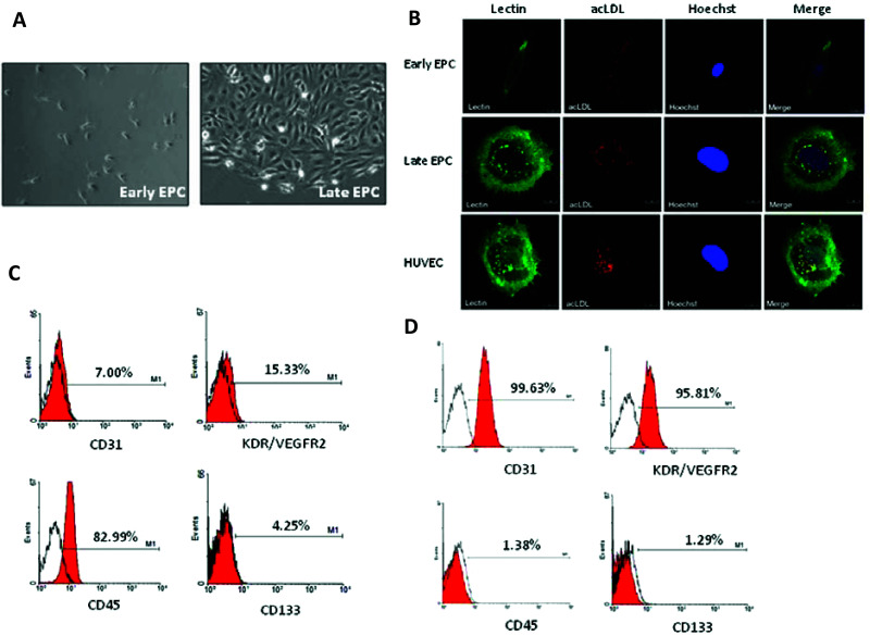
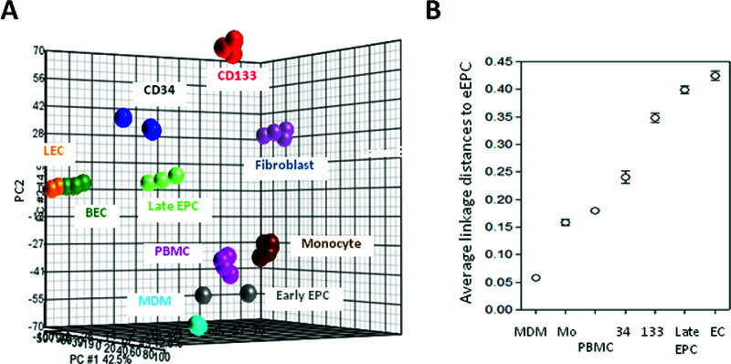
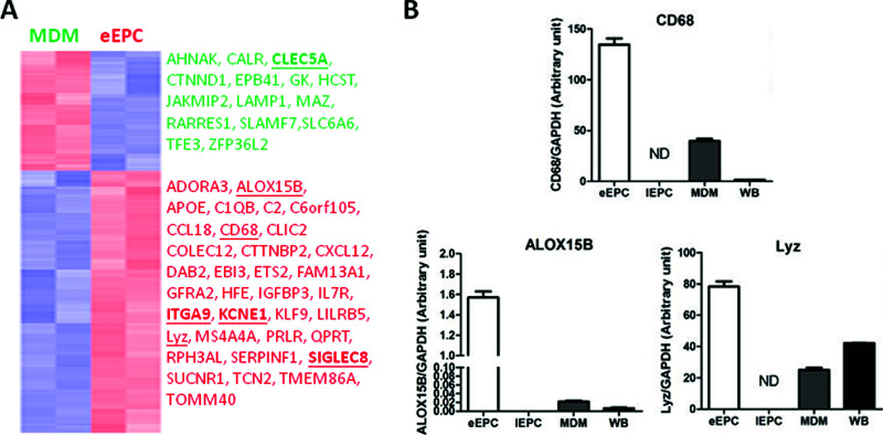
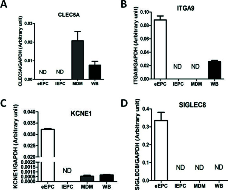
Bone marrow-derived endothelial progenitor cells (EPCs) play a fundamental role in postnatal angiogenesis. Currently, EPCs are defined as early and late EPCs based on their biological properties and their time of appearance during in vitro culture. Reports have shown that early EPCs share common properties and surface markers with adherent blood cells, especially CD14+ monocytes. Distinguishing early EPCs from circulating monocytes or monocyte-derived macrophages (MDMs) is therefore crucial to obtaining pure endothelial populations before they can be applied as part of clinical therapies. We compared the gene expression profiles of early EPCs, blood cells (including peripheral blood mononuclear cells, monocytes, and MDMs), and various endothelial lineage cells (including mature endothelial cells, late EPCs, and CD133+ stem cells). We found that early EPCs expressed an mRNA profile that showed the greatest similarity to MDMs than any other cell type tested. The functional significance of this molecular profiling data was explored by Gene Ontology database search. Novel plasma membrane genes that might potentially be novel isolation biomarkers were also pinpointed. Specifically, expression of CLEC5A was high in MDMs, whereas early EPCs expressed abundant SIGLEC8 and KCNE1. These detailed mRNA expression profiles and the identified functional modules will help to develop novel cell isolation approaches that will allow EPCs to be purified; these can then be used to target cardiovascular disease, tumor angiogenesis, and various ischemia-related diseases.
Conflict of interest statement
The authors declare no conflicts of interests.
Figures
References
-
- Asahara T, Murohara T, Sullivan A, Silver M, van der Zee R, Li T, et al. Isolation of putative progenitor endothelial cells for angiogenesis. Science 1997; 275(5302):964–967. - PubMed
-
- Real C, Caiado F, Dias S. Endothelial progenitors in vascular repair and angiogenesis: How many are needed and what to do? Cardiovasc Hematol Disord Drug Targets 2008; 8(3):185–193. - PubMed
-
- Urbich C, Aicher A, Heeschen C, Dernbach E, Hofmann WK, Zeiher AM, et al. Soluble factors released by endothelial progenitor cells promote migration of endothelial cells and cardiac resident progenitor cells. J Mol Cell Cardiol 2005; 39(5):733–742. - PubMed
-
- Werner N, Kosiol S, Schiegl T, Ahlers P, Walenta K, Link A, et al. Circulating endothelial progenitor cells and cardiovascular outcomes. N Engl J Med 2005; 353(10):999–1007. - PubMed
Publication types
MeSH terms
Substances
LinkOut - more resources
Full Text Sources
Other Literature Sources
Medical
Research Materials