

Tcea3 regulates the vascular differentiation potential of mouse embryonic stem cells
- PMID: 24397209
- PMCID: PMC8750336
- DOI: 10.3727/105221613x13776146743343
Tcea3 regulates the vascular differentiation potential of mouse embryonic stem cells
Abstract
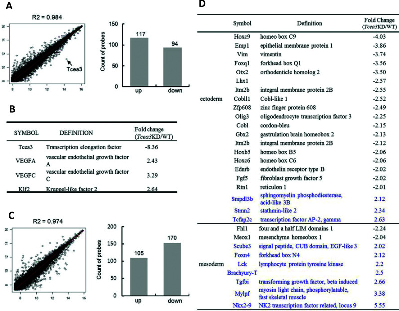
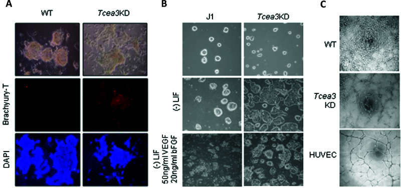
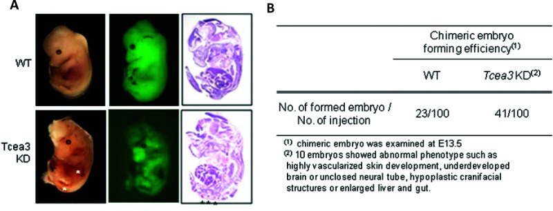
Tcea3 is present in high concentrations in mouse embryonic stem cells (mESCs) and functions to activate Lefly1, a negative regulator of Nodal signaling. The Nodal pathway has numerous biological activities, including mesoderm induction and patterning in early embryogenesis. Here, we demonstrate that the suppression of Tcea3 in mESCs shifts the cells from pluripotency into enhanced mesoderm development. Vascular endothelial growth factor A (VEGFA) and VEGFC, major transcription factors that regulate vasculogenesis, are activated in Tcea3 knocked down (Tcea3 KD) mESCs. Moreover, differentiating Tcea3 KD mESCs have perturbed gene expression profiles with suppressed ectoderm and activated mesoderm lineage markers. Most early differentiating Tcea3 KD cells expressed Brachyury-T, a mesoderm marker, whereas control cells did not express the gene. Finally, development of chimeric embryos that included Tcea3 KD mESCs was perturbed.
Figures
Similar articles
-
Transcription elongation factor Tcea3 regulates the pluripotent differentiation potential of mouse embryonic stem cells via the Lefty1-Nodal-Smad2 pathway.Stem Cells. 2013 Feb;31(2):282-92. doi: 10.1002/stem.1284. Stem Cells. 2013. PMID: 23169579 Free PMC article.
-
TCEA3 promotes differentiation of C2C12 cells via an Annexin A1-mediated transforming growth factor-β signaling pathway.J Cell Physiol. 2019 Jul;234(7):10554-10565. doi: 10.1002/jcp.27726. Epub 2019 Jan 8. J Cell Physiol. 2019. PMID: 30623413
-
Heparan sulfate facilitates FGF and BMP signaling to drive mesoderm differentiation of mouse embryonic stem cells.J Biol Chem. 2012 Jun 29;287(27):22691-700. doi: 10.1074/jbc.M112.368241. Epub 2012 May 3. J Biol Chem. 2012. PMID: 22556407 Free PMC article.
-
Vascular differentiation from embryonic stem cells: novel technologies and therapeutic promises.Vascul Pharmacol. 2012 May-Jun;56(5-6):267-79. doi: 10.1016/j.vph.2012.03.007. Epub 2012 Apr 2. Vascul Pharmacol. 2012. PMID: 22504071 Review.
-
Embryonic stem cell models in vascular biology.J Thromb Haemost. 2009 Jul;7 Suppl 1:53-6. doi: 10.1111/j.1538-7836.2009.03427.x. J Thromb Haemost. 2009. PMID: 19630768 Review.
Cited by
-
Derivation of endothelial cells from porcine induced pluripotent stem cells by optimized single layer culture system.J Vet Sci. 2020 Jan;21(1):e9. doi: 10.4142/jvs.2020.21.e9. J Vet Sci. 2020. PMID: 31940688 Free PMC article.
-
Differentiation of pluripotent stem cells into endothelial cells.Curr Opin Hematol. 2015 May;22(3):252-7. doi: 10.1097/MOH.0000000000000140. Curr Opin Hematol. 2015. PMID: 25767955 Free PMC article. Review.
-
Single-cell Analysis Highlights Anti-apoptotic Subpopulation Promoting Malignant Progression and Predicting Prognosis in Bladder Cancer.Cancer Inform. 2025 Feb 26;24:11769351251323569. doi: 10.1177/11769351251323569. eCollection 2025. Cancer Inform. 2025. PMID: 40018511 Free PMC article.
-
The transcription elongation factor TCEA3 promotes the activity of the myogenic regulatory factors.PLoS One. 2019 Jun 3;14(6):e0217680. doi: 10.1371/journal.pone.0217680. eCollection 2019. PLoS One. 2019. PMID: 31158246 Free PMC article.
-
Gene coexpression networks reveal key drivers of phenotypic divergence in porcine muscle.BMC Genomics. 2015 Feb 5;16(1):50. doi: 10.1186/s12864-015-1238-5. BMC Genomics. 2015. PMID: 25651817 Free PMC article.
References
-
- Evans MJ, Kaufman MH. Establishment in culture of pluripotential cells from mouse embryos. Nature 1981; 292(5819):154–156. - PubMed
-
- Rossant J. Stem cells and early lineage development. Cell 2008, 132(4):527–531. - PubMed
-
- Pera MF, Tam PP. Extrinsic regulation of pluripotent stem cells. Nature 2010; 465(7299):713–720. - PubMed
-
- Watabe T, Miyazono K. Roles of TGF-beta family signaling in stem cell renewal and differentiation. Cell Res 2009; 19(1):103–115. - PubMed
-
- Fei T, Zhu S, Xia K, Zhang J, Li Z, Han JD, et al. Smad2 mediates Activin/Nodal signaling in mesendoderm differentiation of mouse embryonic stem cells. Cell Res 2010; 20(12):1306–1318. - PubMed
Publication types
MeSH terms
Substances
LinkOut - more resources
Full Text Sources
Other Literature Sources
Molecular Biology Databases