. 2014 Apr;224(4):367-76.
doi: 10.1111/joa.12155. Epub 2014 Jan 8.

Midsagittal brain variation and MRI shape analysis of the precuneus in adult individuals

Affiliations

Midsagittal brain variation and MRI shape analysis of the precuneus in adult individuals

Emiliano Bruner et al. J Anat. 2014 Apr.

Abstract

Recent analyses indicate that the precuneus is one of the main centres of integration in terms of functional and structural processes within the human brain. This neuroanatomical element is formed by different subregions, involved in visuo-spatial integration, memory and self-awareness. We analysed the midsagittal brain shape in a sample of adult humans (n = 90) to evidence the patterns of variability and geometrical organization of this area. Interestingly, the major brain covariance pattern within adult humans is strictly associated with the relative proportions of the precuneus. Its morphology displays a marked individual variation, both in terms of geometry (mostly in its longitudinal dimensions) and anatomy (patterns of convolution). No patent differences are evident between males and females, and the allometric effect of size is minimal. However, in terms of morphology, the precuneus does not represent an individual module, being influenced by different neighbouring structures. Taking into consideration the apparent involvement of the precuneus in higher-order human brain functions and evolution, its wide variation further stresses the important role of these deep parietal areas in modern neuroanatomical organization.

Keywords: brain geometry; geometric morphometrics; parietal lobe; shape variation.


Figures

Fig. 1
Fig. 1
The precuneus receives somatosensorial information from the anterior cortex, visual information from the posterior areas, and it is directly connected with the subcortical elements in its inferior boundaries (a) (redrawn after Margulies et al. 2009). In this study, we used 18 landmarks, sampled as 2D coordinates from the midsagittal MRI section (b) (CAS, calcarine sulcus; CC, centre of cerebellum; CG, crista galli; CO, colliculi; GE, genu; IOP, internal occipital protuberance; MB, midbrain; OC, optic chiasm; PCS, postcentral sulcus; POI, parieto-occipital interior; POS, parieto-occipital sulcus; SP, splenium; TC, thalamic centre; sl, sliding landmarks). The limits and average geometry of the precuneus can be visualized by superimposing the whole sample after Procrustes registration of all the landmarks (c) or of only the precuneal area (d). White arrows show the cingulate and perpendicular sulci, the black arrow shows the lower precuneal limits.
Fig. 2
Fig. 2
The standard pattern of circumvolution at the precuneus is an ‘H’ characterized by the subparietal (sp) and precuneal (pc) sulci (images on the left). Such an anatomical organization can be sufficiently recognized in about 60% of the individuals. The rest of the specimens display very different patterns (on the right, some examples). Note also the differences in the patterns of circumvolutions at the junction between calcarine and parieto-occipital sulcus.
Fig. 3
Fig. 3
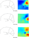
First, second and third principal components of the shape variation for the whole configuration, displayed by wireframes (left) and thin-plate spline deformation grids (right) with deformation maps (red: dilation areas; blue: compression areas).
Fig. 4
Fig. 4
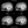
Above: superimposition of the specimens showing the largest and smallest PC1 values (10 individuals for each group), showing the real difference in the relative size of the precuneus between these two extremes of variation. Below: superimposed average images of the whole sample warped according to the PC1 vector, displaying the overall changes of the precuneus proportions along this shape component. In both cases the cingulate sulcus and the parieto-occipital sulcus are easily recognizable, showing the antero-posterior limit of the precuneal area.
Fig. 5
Fig. 5
Larger size involves a longitudinal increase of the precuneus proportions (a), a pattern largely associated with the first principal component. Males have larger parieto-occipital proportions and more dolichocephalic endocranial profile than females (b), although such differences are scanty and probably influenced by brain size. The highest levels of inter-correlation and integration can be observed between the landmarks of the posterior deep areas (c; red links). When analysed alone, the precuneal area shows the first component of variation associated with the longitudinal proportions (d), and the second component associated with the position of contact with the cingulate gyrus (e).

Similar articles

Cited by

References

    1. Ashburner J, Friston KJ. Voxel-based morphometry – The Methods. NeuroImage. 2000;11:805–821. - PubMed
    1. Ashburner J, Friston KJ. Why voxel-based morphometry should be used. NeuroImage. 2001;14:1238–1243. - PubMed
    1. Bastir M, Rosas A, O'Higgins P. Craniofacial levels and the morphological maturation of the human skull. J Anat. 2006;209:637–654. - PMC - PubMed
    1. Bookstein FL. Morphometric Tools for Landmark Data: Geometry and Biology. Cambridge: Cambridge University Press; 1991.
    1. Bookstein FL. “Voxel-based morphometry” should not be used with imperfectly registered images. NeuroImage. 2001;14:1454–1462. - PubMed

Publication types

LinkOut - more resources