

Genetic basis of coaggregation receptor polysaccharide biosynthesis in Streptococcus sanguinis and related species
- PMID: 24397790
- PMCID: PMC3899595
- DOI: 10.1111/omi.12042
Genetic basis of coaggregation receptor polysaccharide biosynthesis in Streptococcus sanguinis and related species
Abstract
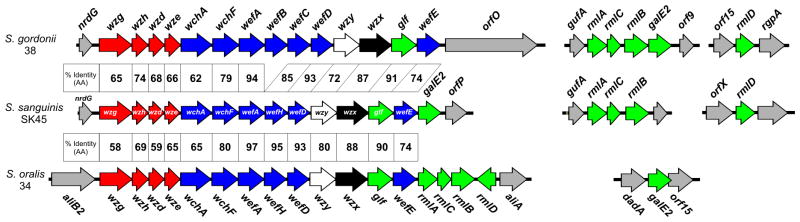
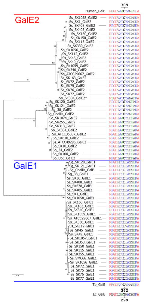
Interbacterial adhesion between streptococci and actinomyces promotes early dental plaque biofilm development. Recognition of coaggregation receptor polysaccharides (RPS) on strains of Streptococcus sanguinis, Streptococcus gordonii and Streptococcus oralis by Actinomyces spp. type 2 fimbriae is the principal mechanism of these interactions. Previous studies of genetic loci for synthesis of RPS (rps) and RPS precursors (rml, galE1 and galE2) in S. gordonii 38 and S. oralis 34 revealed differences between these strains. To determine whether these differences are strain-specific or species-specific, we identified and compared loci for polysaccharide biosynthesis in additional strains of these species and in several strains of the previously unstudied species, S. sanguinis. Genes for synthesis of RPS precursors distinguished the rps loci of different streptococci. Hence, rml genes for synthesis of TDP-L-Rha were in rps loci of S. oralis strains but at other loci in S. gordonii and S. sanguinis. Genes for two distinct galactose epimerases were also distributed differently. Hence, galE1 for epimerization of UDP-Glc and UDP-Gal was in galactose operons of S. gordonii and S. sanguinis strains but surprisingly, this gene was not present in S. oralis. Moreover, galE2 for epimerization of both UDP-Glc and UDP-Gal and UDP-GlcNAc and UDP-GalNAc was at a different locus in each species, including rps operons of S. sanguinis. The findings provide insight into cell surface properties that distinguish different RPS-producing streptococci and open an approach for identifying these bacteria based on the arrangement of genes for synthesis of polysaccharide precursors.
Keywords: Streptococcus sanguinis; biofilm; coaggregation; galactose epimerase; interbacterial adhesion; receptor polysaccharide; rps locus.
Published 2013. This article is a U.S. Government work and is in the public domain in the USA.
Figures
Similar articles
-
Genetic loci for coaggregation receptor polysaccharide biosynthesis in Streptococcus gordonii 38.J Bacteriol. 2003 Sep;185(18):5419-30. doi: 10.1128/JB.185.18.5419-5430.2003. J Bacteriol. 2003. PMID: 12949094 Free PMC article.
-
Coaggregation-mediated interactions of streptococci and actinomyces detected in initial human dental plaque.J Bacteriol. 2003 Jun;185(11):3400-9. doi: 10.1128/JB.185.11.3400-3409.2003. J Bacteriol. 2003. PMID: 12754239 Free PMC article.
-
Characterization of a Streptococcus sp.-Veillonella sp. community micromanipulated from dental plaque.J Bacteriol. 2008 Dec;190(24):8145-54. doi: 10.1128/JB.00983-08. Epub 2008 Sep 19. J Bacteriol. 2008. PMID: 18805978 Free PMC article.
-
Comparative structural and molecular characterization of ribitol-5-phosphate-containing Streptococcus oralis coaggregation receptor polysaccharides.J Bacteriol. 2009 Mar;191(6):1891-900. doi: 10.1128/JB.01532-08. Epub 2009 Jan 16. J Bacteriol. 2009. PMID: 19151140 Free PMC article.
-
Bacteria of healthy periodontal tissues as candidates of probiotics: a systematic review.Eur J Med Res. 2024 Jun 14;29(1):328. doi: 10.1186/s40001-024-01908-2. Eur J Med Res. 2024. PMID: 38877601 Free PMC article.
Cited by
-
Growth Control of Listeria monocytogenes in Raw Sausage via Bacteriocin-Producing Leuconostoc carnosum DH25.Foods. 2024 Jan 17;13(2):298. doi: 10.3390/foods13020298. Foods. 2024. PMID: 38254599 Free PMC article.
-
Mechanistic Effects of E-Liquids on Biofilm Formation and Growth of Oral Commensal Streptococcal Communities: Effect of Flavoring Agents.Dent J (Basel). 2022 May 13;10(5):85. doi: 10.3390/dj10050085. Dent J (Basel). 2022. PMID: 35621538 Free PMC article.
-
Impact of Polymicrobial Infection on Fitness of Streptococcus gordonii In Vivo.mBio. 2023 Jun 27;14(3):e0065823. doi: 10.1128/mbio.00658-23. Epub 2023 Apr 12. mBio. 2023. PMID: 37042761 Free PMC article.
-
Oral streptococci utilize a Siglec-like domain of serine-rich repeat adhesins to preferentially target platelet sialoglycans in human blood.PLoS Pathog. 2014 Dec 4;10(12):e1004540. doi: 10.1371/journal.ppat.1004540. eCollection 2014 Dec. PLoS Pathog. 2014. PMID: 25474103 Free PMC article. Clinical Trial.
-
The dental plaque biofilm matrix.Periodontol 2000. 2021 Jun;86(1):32-56. doi: 10.1111/prd.12361. Epub 2021 Mar 10. Periodontol 2000. 2021. PMID: 33690911 Free PMC article. Review.
References
-
- Bentley SD, Aanensen DM, Mavroidi A, Saunders D, Rabbinowitsch E, Collins M, Donohoe K, Harris D, Murphy L, Quail MA, Samuel G, Skovsted IC, Kaltoft MS, Barrell B, Reeves PR, Parkhill J, Spratt BG. Genetic analysis of the capsular biosynthetic locus from all 90 pneumococcal serotypes. Plos Genet. 2006;2:262–269. - PMC - PubMed
-
- Bergstrom N, Jansson PE, Kilian M, Skov Sorensen UB. Structures of two cell wall-associated polysaccharides of a Streptococcus mitis biovar 1 strain. A unique teichoic acid-like polysaccharide and the group O antigen which is a C-polysaccharide in common with pneumococci. Eur J Biochem. 2000;267:7147–7157. - PubMed
-
- Cisar JO, Sandberg AL, Abeygunawardana C, Reddy GP, Bush CA. Lectin recognition of host-like saccharide motifs in streptococcal cell wall polysaccharides. Glycobiology. 1995;5:655–662. - PubMed
Publication types
MeSH terms
Substances
Associated data
- Actions
- Actions
- Actions
- Actions
- Actions
- Actions
Grants and funding
LinkOut - more resources
Full Text Sources
Other Literature Sources
Miscellaneous
