Case Reports
. 2014 Apr;72(4):702-16.
doi: 10.1016/j.joms.2013.09.008. Epub 2014 Jan 4.

Stage 0 osteonecrosis of the jaw in a patient on denosumab

Affiliations
Case Reports

Stage 0 osteonecrosis of the jaw in a patient on denosumab

Tara L Aghaloo et al. J Oral Maxillofac Surg. 2014 Apr.

Abstract

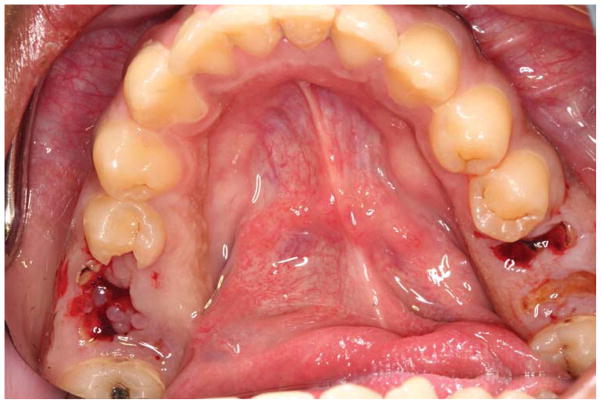
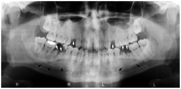
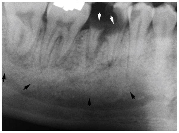
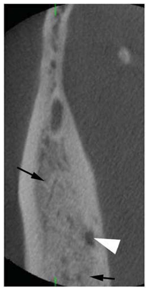
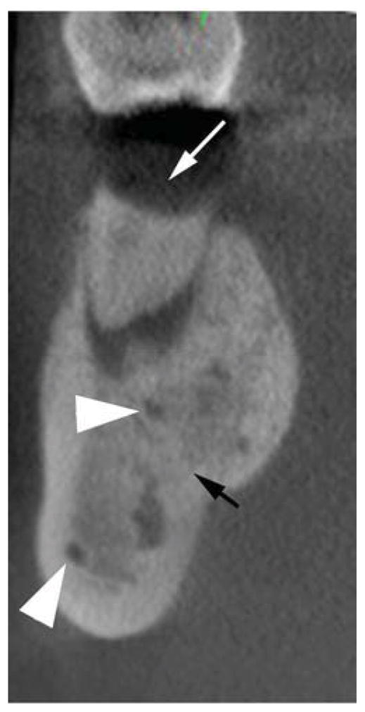
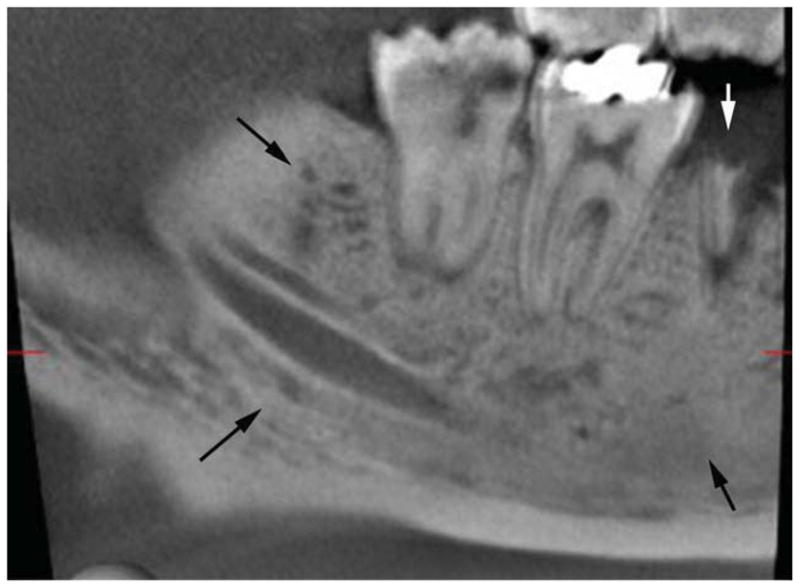
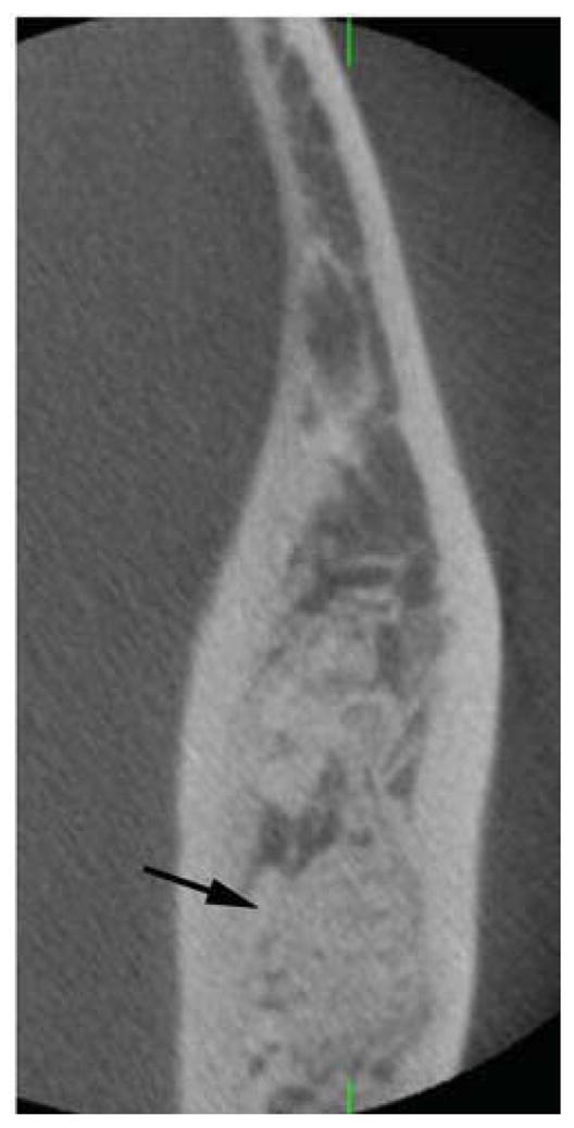
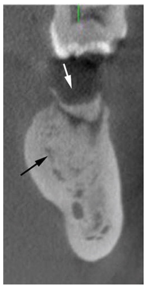
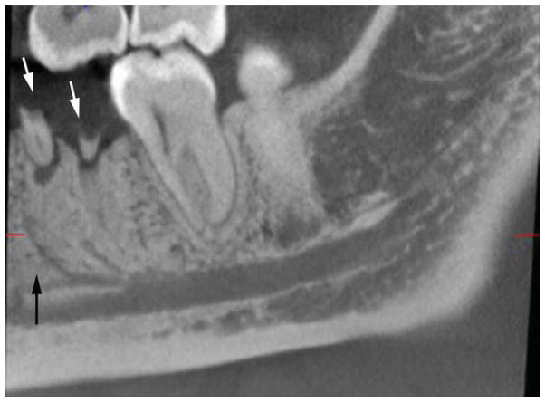
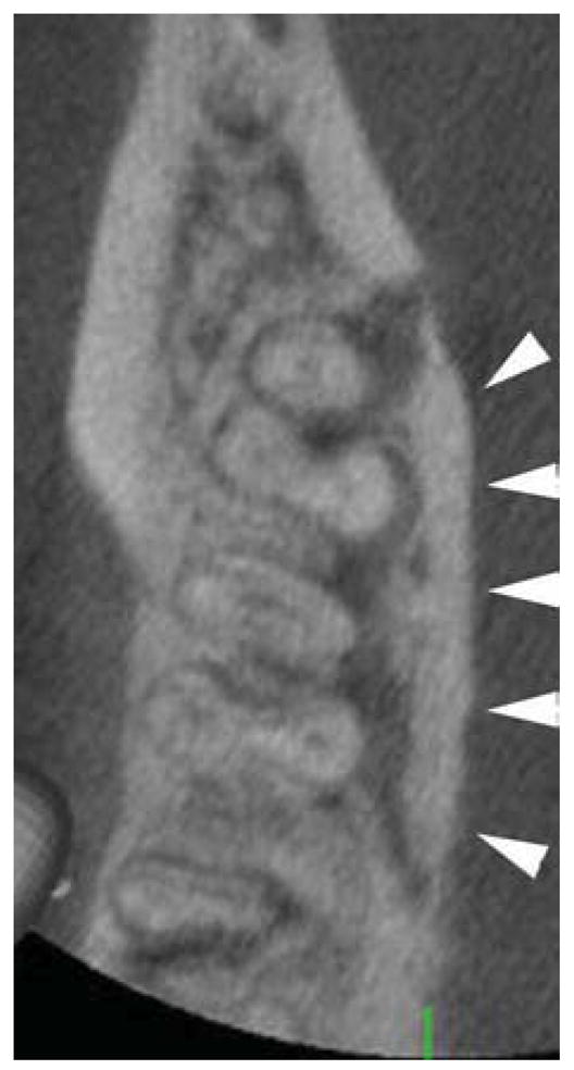
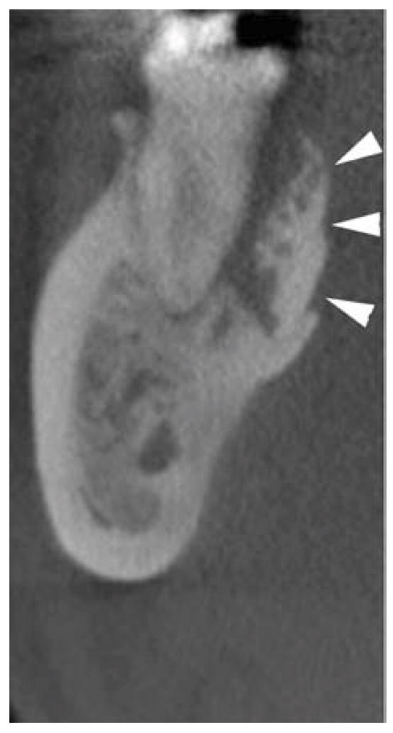
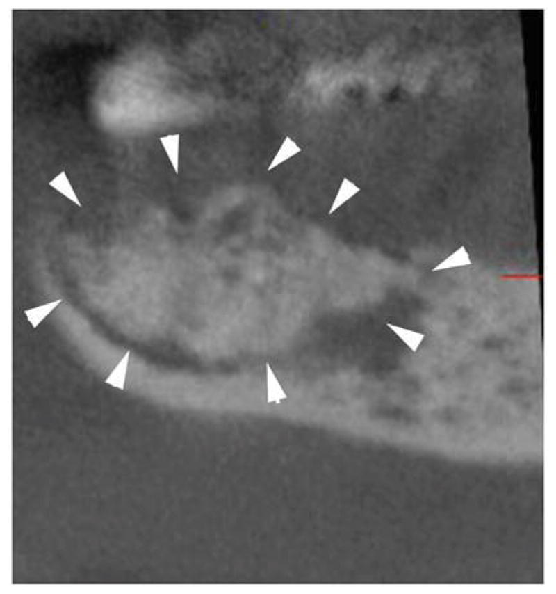
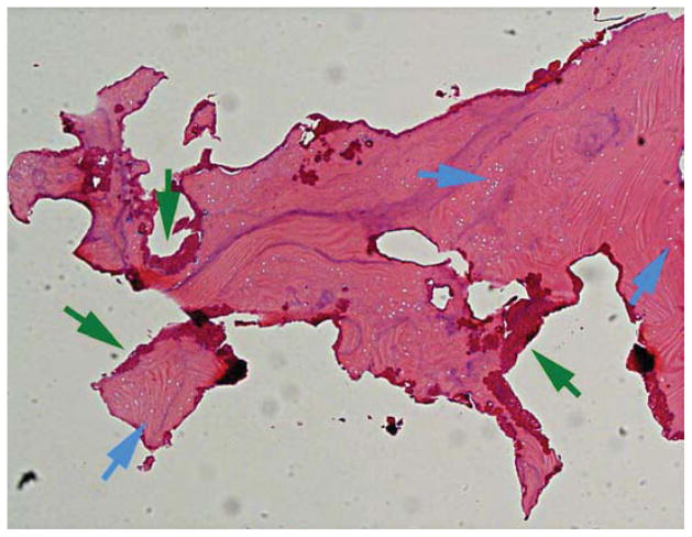
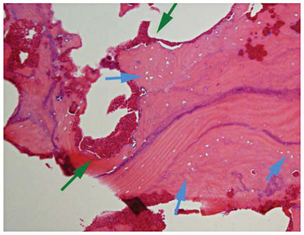
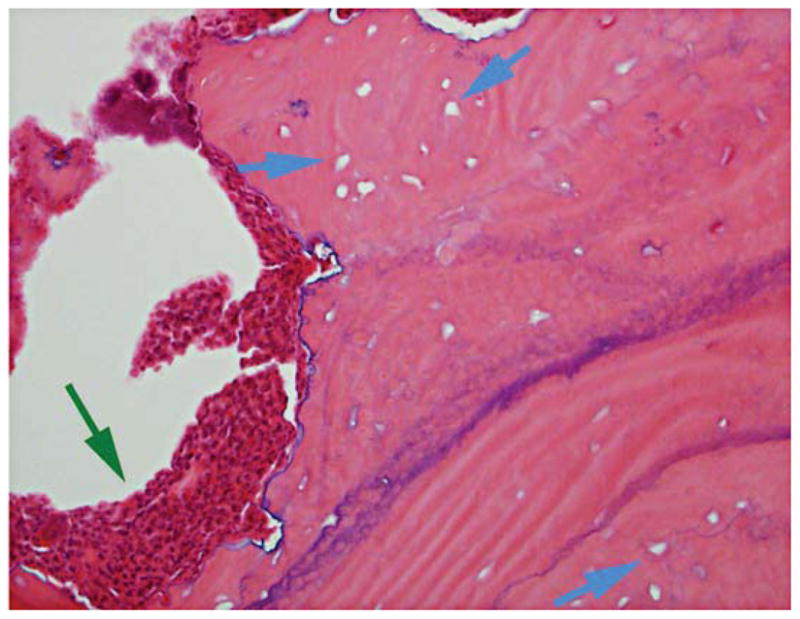
Osteonecrosis of the jaws (ONJ) is a complex disease involving multiple tissue and cell-type responses to wound healing or infection. AAOMS defines bisphosphonate related ONJ (BRONJ) as exposed, necrotic bone in the maxillofacial region that has persisted for more than 8 weeks in a patient with current or previous antiresorptive treatment, without a history of radiation therapy to the jaws. Since the first reported ONJ cases in 2003 and 2004, there has been little advancement in understanding the etiology and pathophysiology of ONJ. Many hypotheses have been proposed, including bisphosphonate (BP) toxicity to oral epithelium, altered wound healing after tooth extraction, high turnover of the mandible and maxilla, oral biofilm formation, infection and inflammation, and suppression of angiogenesis and bone turnover. The current classification system of ONJ involves stages 0 to 3 and is based on patient clinical presentation. This report describes a case of stage 0 ONJ in a patient on denosumab and indicates the full-spectrum similarities between BP- and denosumab-associated ONJ clinically, radiographically, and histologically.


Figures

Figure 1
Figure 1
Clinical presentation of the patient. Root fragments in the areas of #19 and 30 can be seen. However, no evidence of bone exposure or fistula formation is present.
Figure 2
Figure 2
A) Panoramic radiograph of patient at the time of presentation. B) and C) magnified areas of the panoramic radiograph focusing on the posterior right and left mandible, respectively. White arrows point to the root fragments, while black arrows show areas of increased bone density.
Figure 2
Figure 2
A) Panoramic radiograph of patient at the time of presentation. B) and C) magnified areas of the panoramic radiograph focusing on the posterior right and left mandible, respectively. White arrows point to the root fragments, while black arrows show areas of increased bone density.
Figure 2
Figure 2
A) Panoramic radiograph of patient at the time of presentation. B) and C) magnified areas of the panoramic radiograph focusing on the posterior right and left mandible, respectively. White arrows point to the root fragments, while black arrows show areas of increased bone density.
Figure 3
Figure 3
Axial (A, D), cross-sectional (B, E) and sagittal (C, F) cone beam CT (CBCT) slices of the patient’s right (A, B, C) and left (D, E, F) posterior mandible. White arrows point to root fragments, black arrows to areas of increased trabecular density and loss of trabecular architecture, and white arrowhead to radiolucent areas.
Figure 3
Figure 3
Axial (A, D), cross-sectional (B, E) and sagittal (C, F) cone beam CT (CBCT) slices of the patient’s right (A, B, C) and left (D, E, F) posterior mandible. White arrows point to root fragments, black arrows to areas of increased trabecular density and loss of trabecular architecture, and white arrowhead to radiolucent areas.
Figure 4
Figure 4
Axial (A), cross-sectional (B) and sagittal (C) CBCT slices through the area of the lingual cortical plate of the posterior right mandible. White arrowheads delineate the sequestrum outline.
Figure 4
Figure 4
Axial (A), cross-sectional (B) and sagittal (C) CBCT slices through the area of the lingual cortical plate of the posterior right mandible. White arrowheads delineate the sequestrum outline.
Figure 4
Figure 4
Axial (A), cross-sectional (B) and sagittal (C) CBCT slices through the area of the lingual cortical plate of the posterior right mandible. White arrowheads delineate the sequestrum outline.
Figure 5
Figure 5
4X (A), 10X (B), and 20X (C) magnification of H&E stained histologic sections of the bone fragment. Blue arrows point to empty osteocytic lacunae, while green arrows point to neutrophil rimming of the bone. No osteoblasts, osteocytes or osteoclasts are seen.
Figure 5
Figure 5
4X (A), 10X (B), and 20X (C) magnification of H&E stained histologic sections of the bone fragment. Blue arrows point to empty osteocytic lacunae, while green arrows point to neutrophil rimming of the bone. No osteoblasts, osteocytes or osteoclasts are seen.
Figure 5
Figure 5
4X (A), 10X (B), and 20X (C) magnification of H&E stained histologic sections of the bone fragment. Blue arrows point to empty osteocytic lacunae, while green arrows point to neutrophil rimming of the bone. No osteoblasts, osteocytes or osteoclasts are seen.

Similar articles

Cited by

References

    1. Farah CS, Savage NW. Oral ulceration with bone sequestration. Aust Dent J. 2003;48:61. - PubMed
    1. Meer S, Coleman H, Altini M, Alexander T. Mandibular osteomyelitis and tooth exfoliation following zoster-CMV co-infection. Oral Surg Oral Med Oral Pathol Oral Radiol Endod. 2006;101:70. - PubMed
    1. Schwartz HC. Osteonecrosis of the jaws: a complication of cancer chemotherapy. Head Neck Surg. 1982;4:251. - PubMed
    1. Advisory Task Force on Bisphosphonate-Related Ostenonecrosis of the Jaws AAoO, Maxillofacial S American Association of Oral and Maxillofacial Surgeons position paper on bisphosphonate-related osteonecrosis of the jaws. J Oral Maxillofac Surg. 2007;65:369. - PubMed
    1. Ruggiero SL, Dodson TB, Assael LA, Landesberg R, Marx RE, Mehrotra B American Association of O Maxillofacial S. American Association of Oral and Maxillofacial Surgeons position paper on bisphosphonate-related osteonecrosis of the jaws--2009 update. J Oral Maxillofac Surg. 2009;67:2. - PubMed

Publication types

MeSH terms