. 2014 Jan 7;16(1):R1.
doi: 10.1186/bcr3593.

New insights into lineage restriction of mammary gland epithelium using parity-identified mammary epithelial cells

New insights into lineage restriction of mammary gland epithelium using parity-identified mammary epithelial cells

Ted H-T Chang et al. Breast Cancer Res. .

Abstract

Introduction: Parity-identified mammary epithelial cells (PI-MECs) are an interesting cellular subset because they survive involution and are a presumptive target for transformation by human epidermal growth factor receptor 2 (HER2)/neu in mammary tumors. Depending on the type of assay, PI-MECs have been designated lobule-restricted progenitors or multipotent stem/progenitor cells. PI-MECs were reported to be part of the basal population of mammary epithelium based on flow cytometry. We investigated the cellular identity and lineage potential of PI-MECs in intact mammary glands.

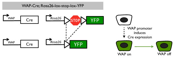
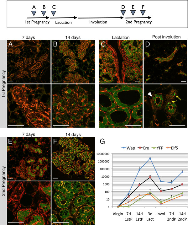
Methods: We performed a quantitative and qualitative analysis of the contribution of PI-MECs to mammary epithelial cell lineages in pregnant and involuted mammary glands by immunohistochemistry, fluorescence-activated cells sorting (FACS), and quantitative polymerase chain reaction. PI-MECs were labeled by the activation of Whey Acidic Protein (WAP)-Cre during pregnancy that results in permanent expression of yellow fluorescent protein.

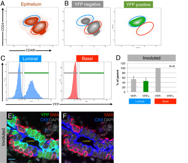
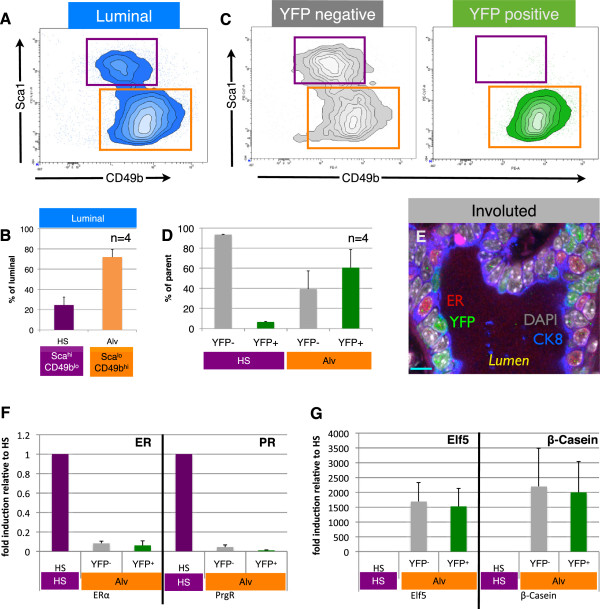
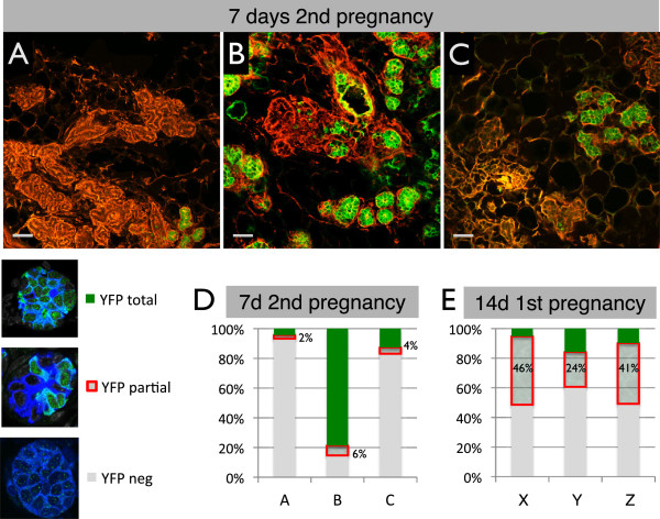
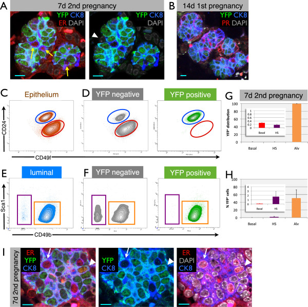
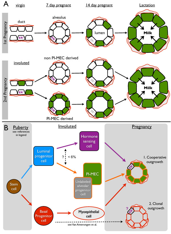
Results: After involution, PI-MECs are present exclusively in the luminal layer of mammary ducts. During pregnancy, PI-MECs contribute to the luminal layer but not the basal layer of alveolar lobules. Strikingly, whereas all luminal estrogen receptor (ER)-negative cells in an alveolus can be derived from PI-MECs, the alveolar ER-positive cells are unlabeled and reminiscent of Notch2-traced L cells. Notably, we observed a significant population of unlabeled alveolar progenitors that resemble PI-MECs based on transcriptional and histological analysis.

Conclusions: Our demonstration that PI-MECs are luminal cells underscores that not only basal cells display multi-lineage potential in transplantation assays. However, the lineage potential of PI-MECs in unperturbed mammary glands is remarkably restricted to luminal ER-negative cells of the secretory alveolar lineage. The identification of an unlabeled but functionally similar population of luminal alveolar progenitor cells raises the question of whether PI-MECs are a unique population or the result of stochastic labeling. Interestingly, even when all luminal ER-negative cells of an alveolus are PI-MEC-derived, the basal cells and hormone-sensing cells are derived from a different source, indicating that cooperative outgrowth of cells from different lineages is common in alveologenesis.


Figures

Figure 1
Figure 1
Labeling of parity-identified mammary epithelial cells (PI-MECs). In WAP-Cre;Rosa26-lsl-YFP double transgenic mice, yellow fluorescent protein (YFP) is not expressed (white cell) until the whey acidic protein (WAP) promoter is induced (by pregnancy). When activation of the WAP promoter results in the expression of sufficient levels of the Cre recombinase, the loxP sites (triangles) will be recombined and the sequence in between will be excised (dotted line), bringing the YFP gene under control of the constitutively active Rosa26 promoter (green cell, WAP on). Upon cell division, daughter cells will remain YFP-positive even though WAP is not expressed (green cell, WAP off) as a result of the genetic deletion of the stop sequence.
Figure 2
Figure 2
Analysis of parity-identified mammary epithelial cells (PI-MECs) during first and second pregnancies. Mammary glands from WAP-Cre; Rosa-lsl-YFP mice were collected at the following stages: 7 days (A) and 14 days (B) of the first pregnancy, 3 days of lactation (C), involuted (>6 weeks after weaning) (D), and 7 days (E) and 14 days (F) of the second pregnancy. Cryosections were analyzed for YFP expression (green) and counterstained with rhodamine-conjugated phalloidin (red). Representative confocal microscope images were captured with 20× (upper panels) and 63× (lower panels) objective lenses. Bar is 50 μm. White arrowhead indicates a PI-MEC (YFPpos cell that survived involution), and the yellow arrows indicate collapsed YFP bodies seen often in involuted sections. (G) Quantification of relative mRNA levels in mammary epithelial organoid isolates. Gene expression is normalized to hypoxanthine-guanine phosphoribosyltransferase (HPRT) expression, and error bars indicate standard deviation of three individual mice at each time point.
Figure 3
Figure 3
Parity-identified mammary epithelial cells (PI-MECs) in involuted mammary glands are luminal. (A) Epithelial cells isolated from involuted mammary glands were labeled with fluorescent antibodies against CD24 and α6-integrin (CD49f) to distinguish the luminal (blue gate) and basal (red gate) population. (B) Yellow fluorescent protein-negative (YFPneg) cells and YFPpos cells (PI-MECs) are shown separately on density/contour plots. (C) The luminal and basal populations are plotted in separate histograms to quantify the proportion of YFP-positive cells in each population (D); error bars indicate standard deviation for 4 individual mice. (E) Immunofluorescent probing of involuted mammary epithelium for YFP (PI-MECs, green), the luminal marker cytokeratin-8 (CK8) (blue), and basal marker smooth muscle actin (SMA) (red). Nuclei are labeled by 4′,6-diamidino-2-phenylindole (DAPI) (grey). (F) Omission of the YFP signal from the image shown in (E) shows the mutual exclusive labeling of luminal cells by CK8 (blue) and basal cells by SMA (red). Bar is 10 μm.
Figure 4
Figure 4
Parity-identified mammary epithelial cells (PI-MECs) in involuted mammary glands belong to the alveolar progenitor population. (A) The luminal population of mammary epithelial cells (CD24hi CD49flo) is separated into hormone-sensing cells (Sca1hi CD49blo; purple gate) and alveolar progenitor cells (Sca1lo CD49bhi; orange gate). (B) Relative proportions of hormone-sensing (HS) cells and alveolar progenitor cells (Alv) within the luminal population. (C) Analysis of yellow fluorescent protein-negative (YFPneg) (grey) and YFPpos (PI-MEC, green) subpopulations of luminal cells shows that PI-MECs are found mainly in the alveolar progenitor cell population (orange gate). Note that the combined density/contour plot shows the relative distribution of the population on display and therefore the HS population appears larger in (C) than in (A) because the YFPpos population is plotted separately in (C). (D) Distribution of YFPneg cells and YFPpos PI-MECs within populations of HS cells (Sca1hi CD49blo; purple label) and of alveolar progenitor cells (Sca1lo CD49bhi; orange label). (E) Immunofluorescent probing of an involuted mammary section identifies three luminal cell types: HS cells expressing the estrogen receptor alpha (ERα) (red), PI-MECs expressing YFP (green), and luminal cells expressing CK8 (blue) but neither ER nor YFP. Bar is 10 μm. (F) Relative mRNA expression levels of HS cell marker genes ERα and progesterone receptor (PR) and (G) alveolar cell marker genes Elf5 and β-Casein on populations of sorted HS cells (Sca1hiCD49blo) or alveolar progenitor cells (Sca1loCD49bhi) which were separated based on their YFP expression. Error bars indicate standard deviation for four individual mice.
Figure 5
Figure 5
A large proportion of alveoli derive from non-parity-identified mammary epithelial cells (non-PI-MECs) during the second pregnancy. (A-C) Cryosections stained with phalloidin (red) of three independent mice used to quantify the relative contribution of PI-MECs to alveolar development at day 7 of the second pregnancy. Scale bar is 50 μm. (D) The vast majority of alveoli were scored as either completely yellow fluorescent protein (YFP)-negative (grey) or completely YFP-positive (green), with few alveoli containing a mixture of the two, as highlighted by the red box. (E) Quantification of the YFP pattern in alveoli of three mice (X-Z) at day 14 of the first pregnancy (a time of de novo floxing). At least 750 alveoli/mouse were counted.
Figure 6
Figure 6
Parity-identified mammary epithelial cells (PI-MECs) remain lineage-restricted during pregnancy. (A) Immunofluorescence on paraffin sections from the 7-day second pregnancy time point, showing alveoli in which all cells are yellow fluorescent protein-positive (YFPpos), except for rare luminal cells expressing estrogen receptor alpha (ERα) (red, yellow arrows) or basal (CK8-negative, white arrowhead) cells. (B) Alveolar clusters at day 14 of the first pregnancy that already consist mostly of YFPpos cells also contain hormone-sensing (HS) cells (labeled by an antibody against the progesterone receptor, red) which do not express YFP. Scale bar is 10 μm. (C) Mammary epithelial cells from 7-day pregnant glands (second pregnancy) labeled with CD24 and CD49f to segregate luminal (blue gate) from basal (red gate) single-cell populations. (D) YFPneg cells and YFPpos cells (PI-MECs) are shown separately on density/contour plots for CD24 and CD49f. (E) Sca1 and CD49b plots of the luminal cell population at 7 days of pregnancy, showing the HS cell population (purple gate) and alveolar cells (orange gate). (F) Distribution of YFPneg cells and YFPpos PI-MECs within populations of HS cells and of alveolar cells. Note that the combined density/contour plot shows the relative distribution of the population on display and therefore the HS population appears larger in (D) than in (C) because the YFPpos population is plotted separately in (D). (G) Distribution of YFPpos cells across the different populations. Inset: magnified basal and HS cell bars. (H) Percentage of YFPpos cells observed within the basal, HS, and alveolar cell populations. Error bars indicate standard deviation of four independent mice. (I) A small percentage of ER+ cells are also YFPpos (arrowhead), although most HS cells are YFPneg (arrow). Scale bar is 10 μm.
Figure 7
Figure 7
PI-MEC lineage tracing (A) and a stem cell hierarchy for intact mammary glands (B). (A) The YFP reporter becomes robustly activated by 14 days of the first pregnancy (green) and by lactation virtually all secretory cells are labeled. After involution, labeled cells are present in the luminal layer of the ducts (only stage where PI-MECs can be definitively recognized, black diamond). At 7 days of the second pregnancy, PI-MECs give rise to all of the ER-negative luminal cells of some but not all alveoli (WAP promoter activity is low at this stage). PI-MECs do not generate the alveolar basal cells (red) and ER+ cells (purple). By 14 days of the second pregnancy, the WAP promoter is significantly induced, and ER-negative luminal cells in alveoli become labeled de novo. ER expression becomes more difficult to detect (grey), but the hormone-sensing cells still express PR and not YFP (see Figure 6). (B) Bipotent stem cell activity was reported during embryonic development and puberty [10,34]. In adult non-pregnant mammary glands, unipotent cells maintain the luminal and basal layer [10,34]. We show that PI-MECs are located in the luminal layer and belong to the alveolar lineage (orange). This lineage also contains a population of unlabeled cells with a similar cell surface and gene expression profile. A small proportion of ER+ cells is also YFPpos (< 6%) but it is unclear whether this is due to lineage plasticity or a technical artifact. During pregnancy, a significant proportion of alveoli is formed by cooperative outgrowth of ER-negative luminal cells , hormone-sensing cells and basal cells (7B-1, see Figure 6 and Discussion). An unknown proportion of alveoli contain luminal and basal cells derived from the same Axin2-traced basal progenitor [34] (7B-2), it remains to be tested if this progenitor also generates hormone-sensing cells.

Similar articles

Cited by

References

    1. Visvader JE. Keeping abreast of the mammary epithelial hierarchy and breast tumorigenesis. Genes Dev. 2009;23:2563–2577. doi: 10.1101/gad.1849509. - DOI - PMC - PubMed
    1. Regan JL, Sourisseau T, Soady K, Kendrick H, McCarthy A, Tang C, Brennan K, Linardopoulos S, White DE, Smalley MJ. Aurora a kinase regulates mammary epithelial cell fate by determining mitotic spindle orientation in a notch-dependent manner. Cell Rep. 2013;4:110–123. doi: 10.1016/j.celrep.2013.05.044. - DOI - PubMed
    1. Kordon E, Smith G. An entire functional mammary gland may comprise the progeny from a single cell. Development. 1998;125:1921–1930. - PubMed
    1. Stingl J, Eaves C, Zandieh I, Emerman J. Characterization of bipotent mammary epithelial progenitor cells in normal adult human breast tissue. Breast Cancer Res Treat. 2001;67:93–109. doi: 10.1023/A:1010615124301. - DOI - PubMed
    1. Perou CM, Sørlie T, Eisen MB, van de Rijn M, Jeffrey SS, Rees CA, Pollack JR, Ross DT, Johnsen H, Akslen LA, Fluge O, Pergamenschikov A, Williams C, Zhu SX, Lønning PE, Børresen-Dale AL, Brown PO, Botstein D. Molecular portraits of human breast tumours. Nature. 2000;406:747–752. doi: 10.1038/35021093. - DOI - PubMed

Publication types

MeSH terms