

Ovarian lymphoma and hydronephrosis
- PMID: 24398216
- PMCID: PMC3866078
- DOI: 10.4293/108680813X13654754534918
Ovarian lymphoma and hydronephrosis
Abstract
Introduction: Ovarian lymphoma is a rare entity, and hydronephrosis from lymphoma is even rarer. Most reports describe a laparoscopic approach to the disease, but we report a case of hydroureteronephrosis associated with ovarian lymphoma managed completely by miniinvasive techniques.
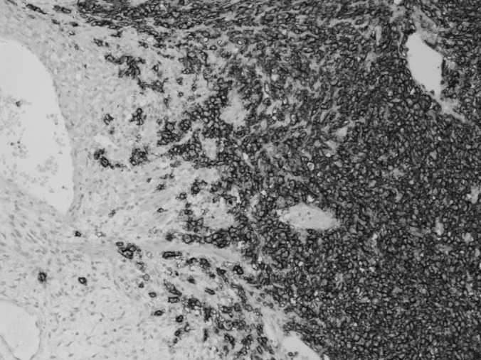
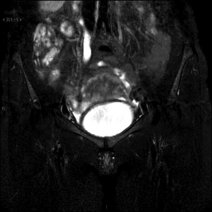
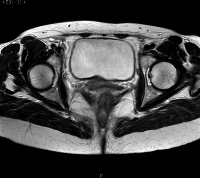
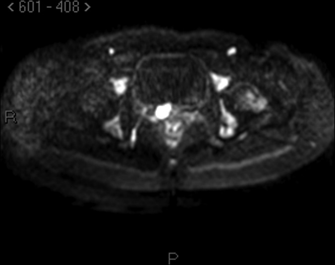
Case report: A 51-year-old woman was referred to us for back pain and renal colic and computed tomography scan findings of right hydroureteronephrosis and a mass in the right mesorectum and uterosacral ligament. After magnetic resonance imaging was performed, the patient underwent laparoscopic adnexectomy and ureterolysis after ureteroscopy and stenting. Histology results showed diffuse B-cell lymphoma of the ovary occluding the ureter without infiltration. The patient has undergone 6 cycles of chemotherapy.
Discussion: This is the first report to describe ovarian lymphoma and hydroureteronephrosis managed completely by laparoscopic surgery and endoscopy. Frequency in clinical practice, differential diagnosis, and endoscopic approach are discussed. The advantages of a multidisciplinary endoscopic team are underlined.
Figures
Similar articles
-
A rare cause of hydronephrosis: Leiomyoma of the ureter and a literature review.Urologia. 2023 Feb;90(1):189-191. doi: 10.1177/03915603211010463. Epub 2021 Apr 19. Urologia. 2023. PMID: 33870770 Review.
-
Retroperitoneal laparoscopy management for ureteral fibroepithelial polyps causing hydronephrosis in children: a report of five cases.J Pediatr Urol. 2015 Oct;11(5):257.e1-5. doi: 10.1016/j.jpurol.2015.02.019. Epub 2015 Apr 24. J Pediatr Urol. 2015. PMID: 25982337
-
Robot-Assisted Laparoscopic Excision of Ureteral and Ureteropelvic Junction Fibroepithelial Polyps in Children.J Endourol. 2016 Aug;30(8):896-900. doi: 10.1089/end.2016.0006. J Endourol. 2016. PMID: 27279495
-
Deep infiltrating ureteral endometriosis with catamenial hydroureteronephrosis: a case report.J Med Case Rep. 2017 Dec 13;11(1):346. doi: 10.1186/s13256-017-1518-6. J Med Case Rep. 2017. PMID: 29233171 Free PMC article.
-
[Bilateral multiple ureteral polyps causing intermittent hydronephrosis: a case report].Nihon Hinyokika Gakkai Zasshi. 2008 Jan;99(1):43-7. doi: 10.5980/jpnjurol1989.99.43. Nihon Hinyokika Gakkai Zasshi. 2008. PMID: 18260348 Review. Japanese.
Cited by
-
Complications of lymphoma in the abdomen and pelvis: clinical and imaging review.Abdom Radiol (NY). 2022 Aug;47(8):2937-2955. doi: 10.1007/s00261-022-03567-5. Epub 2022 Jun 12. Abdom Radiol (NY). 2022. PMID: 35690955 Free PMC article. Review.
References
-
- Signorelli M, Maneo A, Cammarota S, et al. Conservative management in primary genital lymphomas: the role of chemottherapy. Gynecol Oncol. 2007;104:416–423 - PubMed
-
- Monterroso V, Jaffe ES, Merino MJ, Medeiros LJ. Malignant lymphomas involving the ovary. A clinicopathologic analysis of 39 cases. Am J Surg Pathol. 1993;17:154–170 - PubMed
-
- Dimopoulos MA, Daliani D, Pugh W, Gershenson D, Cabanillas F, Sarris AH. Primary ovarian non-Hodgkin's lymphoma: outcome after treatment with combination chemotherapy. Gynecol Oncol. 1997;64:446–450 - PubMed
-
- Bambury I, Wharfe G, Fletcher H, Williams E, Jaggon J. Ovarian lymphoma. J Obstet Gynecol. 2011;31:653–655 - PubMed
Publication types
MeSH terms
LinkOut - more resources
Full Text Sources
Other Literature Sources
Medical