

A recurrent 11q aberration pattern characterizes a subset of MYC-negative high-grade B-cell lymphomas resembling Burkitt lymphoma
- PMID: 24398325
- PMCID: PMC3931189
- DOI: 10.1182/blood-2013-06-507996
A recurrent 11q aberration pattern characterizes a subset of MYC-negative high-grade B-cell lymphomas resembling Burkitt lymphoma
Abstract
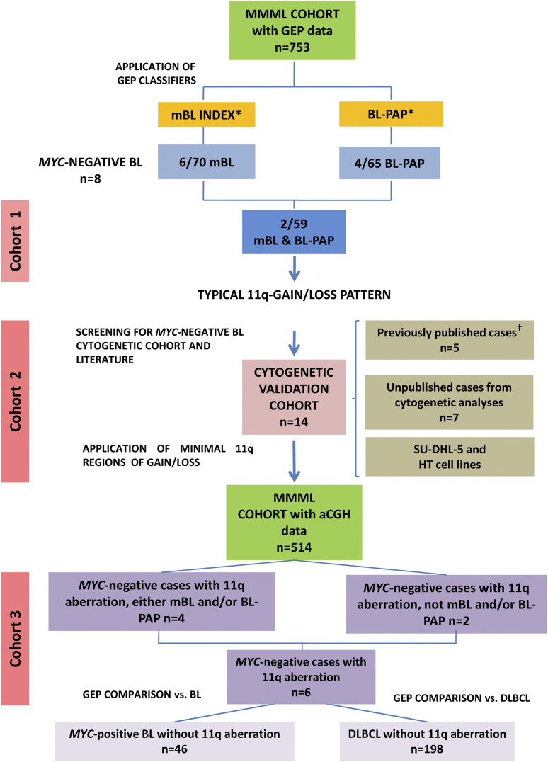
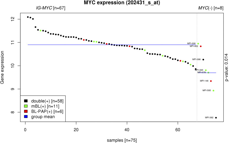
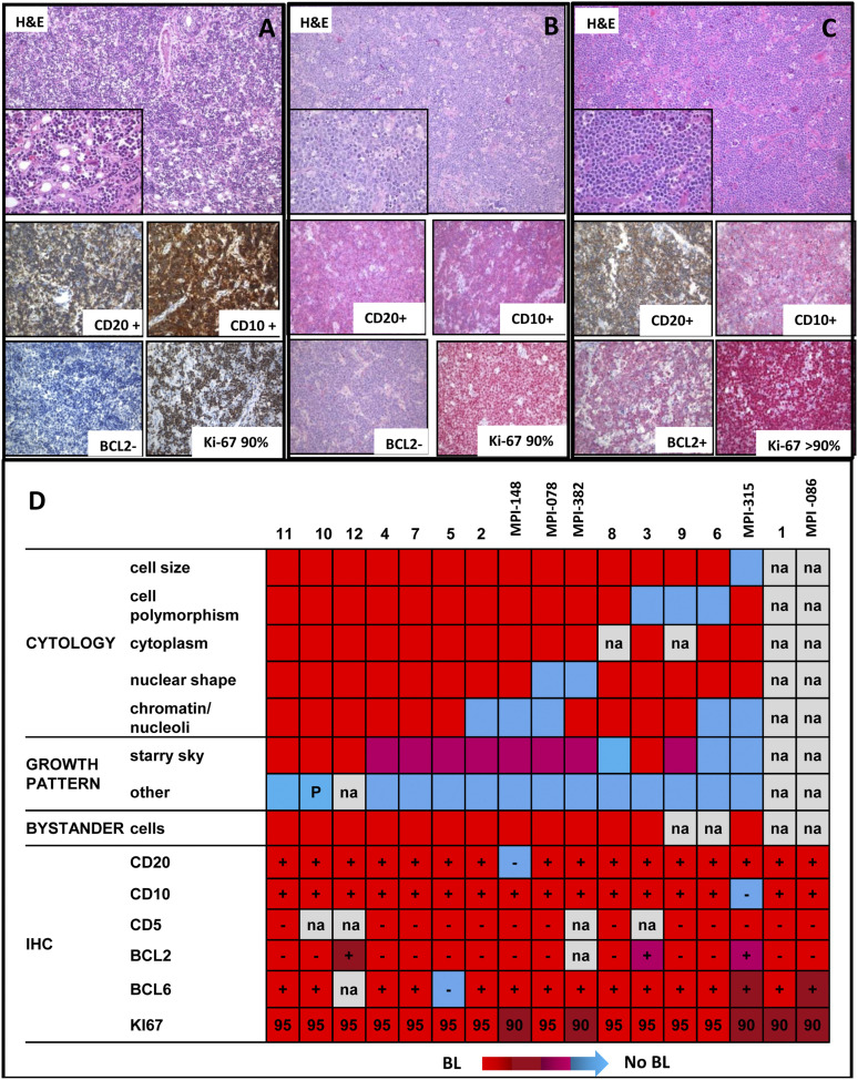
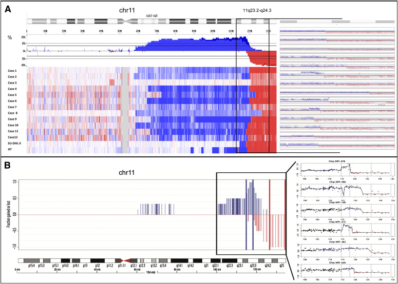
The genetic hallmark of Burkitt lymphoma (BL) is the t(8;14)(q24;q32) and its variants leading to activation of the MYC oncogene. It is a matter of debate whether true BL without MYC translocation exists. Here, we identified 59 lymphomas concordantly called BL by 2 gene expression classifiers among 753 B-cell lymphomas. Only 2 (3%) of these 59 molecular BL lacked a MYC translocation, which both shared a peculiar pattern of chromosome 11q aberration characterized by interstitial gains including 11q23.2-q23.3 and telomeric losses of 11q24.1-qter. We extended our analysis to 17 MYC-negative high-grade B-cell lymphomas with a similar 11q aberration and showed this aberration to be recurrently associated with morphologic and clinical features of BL. The minimal region of gain was defined by high-level amplifications in 11q23.3 and associated with overexpression of genes including PAFAH1B2 on a transcriptional and protein level. The recurrent region of loss contained a focal homozygous deletion in 11q24.2-q24.3 including the ETS1 gene, which was shown to be mutated in 4 of 16 investigated cases. These findings indicate the existence of a molecularly distinct subset of B-cell lymphomas reminiscent of BL, which is characterized by deregulation of genes in 11q.
Figures
References
-
- Swerdlow S, Campo E, Harris N, et al., editors. WHO Classification of Tumours of Haematopoietic and Lymphoid Tissues. IARC Lyon: 2008.
-
- Boxer LM, Dang CV. Translocations involving c-myc and c-myc function. Oncogene. 2001;20(40):5595–5610. - PubMed
-
- Hecht JL, Aster JC. Molecular biology of Burkitt’s lymphoma. J Clin Oncol. 2000;18(21):3707–3721. - PubMed
-
- Salaverria I, Siebert R. The gray zone between Burkitt’s lymphoma and diffuse large B-cell lymphoma from a genetics perspective. J Clin Oncol. 2011;29(14):1835–1843. - PubMed
-
- Leucci E, Cocco M, Onnis A, et al. MYC translocation-negative classical Burkitt lymphoma cases: an alternative pathogenetic mechanism involving miRNA deregulation. J Pathol. 2008;216(4):440–450. - PubMed
Publication types
MeSH terms
LinkOut - more resources
Full Text Sources
Other Literature Sources
Molecular Biology Databases
Miscellaneous
