. 2014 Jan;6(1):213-27.
doi: 10.1093/gbe/evu006.

Molecular evolutionary characterization of a V1R subfamily unique to strepsirrhine primates

Affiliations

Molecular evolutionary characterization of a V1R subfamily unique to strepsirrhine primates

Anne D Yoder et al. Genome Biol Evol. 2014 Jan.

Abstract

Vomeronasal receptor genes have frequently been invoked as integral to the establishment and maintenance of species boundaries among mammals due to the elaborate one-to-one correspondence between semiochemical signals and neuronal sensory inputs. Here, we report the most extensive sample of vomeronasal receptor class 1 (V1R) sequences ever generated for a diverse yet phylogenetically coherent group of mammals, the tooth-combed primates (suborder Strepsirrhini). Phylogenetic analysis confirms our intensive sampling from a single V1R subfamily, apparently unique to the strepsirrhine primates. We designate this subfamily as V1Rstrep. The subfamily retains extensive repertoires of gene copies that descend from an ancestral gene duplication that appears to have occurred prior to the diversification of all lemuriform primates excluding the basal genus Daubentonia (the aye-aye). We refer to the descendent clades as V1Rstrep-α and V1Rstrep-β. Comparison of the two clades reveals different amino acid compositions corresponding to the predicted ligand-binding site and thus potentially to altered functional profiles between the two. In agreement with previous studies of the mouse lemur (genus, Microcebus), the majority of V1Rstrep gene copies appear to be intact and under strong positive selection, particularly within transmembrane regions. Finally, despite the surprisingly high number of gene copies identified in this study, it is nonetheless probable that V1R diversity remains underestimated in these nonmodel primates and that complete characterization will be limited until high-coverage assembled genomes are available.

Keywords: G-protein-coupled receptors; chemosensory genes; gene family evolution; lemurs; olfaction; positive selection.


Figures

F<sc>ig</sc>. 1.—
Fig. 1.—
Time-scaled phylogeny for strepsirrhine primates included in this study. Data and 95% credible divergence ages were derived from Horvath et al. (2008). Some nodes do not show age estimates as not all species examined in this study were included in the Horvath et al. (2008) analysis. Branches are colored as follows: Lorisiformes (red), Daubentoniidae (orange), Indriidae (yellow), Cheirogaleidae (blue), and Lemuridae (green). First column on right indicates whether urine washing is practiced by that species. Second column indicates whether the species is nocturnal (N), diurnal (D), or cathemeral (C). Behavioral data were taken from Delbarco-Trillo et al. (2011). Latin binomials with common names of species are O. garnettii (small-eared galago), Galago moholi (mohol bushbaby), Nycticebus pygmaeus (pygmy slow loris), Daubentonia madagascariensis (aye-aye), Propithecus coquereli (Coquerel's sifaka), Propithecus tattersalli (Tattersall's sifaka), Phaner pallescens (pale fork-marked lemur), Cheirogaleus medius (fat-tailed dwarf lemur), C. major (greater dwarf lemur), Allocebus trichotis (hairy-eared dwarf lemur), Microcebus simmonsi (Simmon's mouse lemur), M. murinus (gray mouse lemur), M. griseorufus (gray–brown mouse lemur), Varecia rubra (red ruffed lemur), V. variegata (black and white ruffed lemur), Hapalemur griseus (eastern lesser bamboo lemur), Lemur catta (ring-tailed lemur), Eulemur mongoz (mongoose lemur), and Eulemur collaris (collared brown lemur).
F<sc>ig</sc>. 2.—
Fig. 2.—
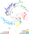
RAxML maximum likelihood tree of V1R sequences generated herein combined with a subset of those identified by Young et al. (2010). (A) V1R sequences originating from strepsirrhine primates (lemurs and lorises) are shown in red. The largest red clade corresponds to the V1Rstrep subfamily. (B) Primate V1R sequences: lemuriforms (blue); lorisiforms (red); tarsiers (yellow); and anthropoids (green).
F<sc>ig</sc>. 3.—
Fig. 3.—
Maximum likelihood phylogeny based on DNA sequence data showing the relationships among V1Rstrep gene copies with the α and β clades indicated. Red represents the lorisiform primates sampled herein (Otolemur, Galago, and Nycticebus), orange represents family Daubentoniidae (genus Daubentonia), green indicates the family Lemuridae (genera Lemur, Hapalemur, Eulemur, and Varecia), yellow indicates family Indriidae (genus Propithecus), and blue represents family Cheirogaleidae (genera Microcebus, Cheirogaleus, Phaner, and Allocebus). It is notable that lemurids (green) are far more abundant in the α clade than in the β clade; conversely, indriids (yellow) are more abundant in the β clade than in the α clade. For precise taxonomic identities represented, refer to supplementary table S1, Supplementary Material online.
F<sc>ig</sc>. 4.—
Fig. 4.—
Maximum likelihood phylogenies based on clustered DNA sequence data (98% similarity threshold) of the V1Rstrep α and β repertoires for Cheirogaleidae and Lemuridae. Phylogenetic analyses were performed on centroid sequences of clusters sharing less than 98% similarity. Results of RaxML analyses on combined (A) and family specific analyses of Cheirogaleidae (B) and Lemuridae (C) are shown. Nodal support was measured by performing 500 bootstrap iterations using RaxML and PhyML (top percentages, respectively) and with Bayesian posterior probabilities based on 2 million iterations (bottom score). Daubentonia was used as the outgroup for rooting both trees (not shown). NS, no support in all three phylogenetic methods.
F<sc>ig</sc>. 5.—
Fig. 5.—
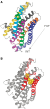
Predicted protein structure and function for amino acid sequences in our alignment using I-TASSER (http://zhanglab.ccmb.med.umich.edu/I-TASSER/, last accessed January 17, 2014). (A) Model of a seven TM G protein-coupled receptor: yellow, TM1; light blue, TM2; green, TM3; purple, TM4; dark blue, TM5; orange, TM6; and dark gray, TM7. Arrow identifies the orientation of the protein with respect to extracellular (EXT) and intracellular (INT) regions. (B) I-TASSER predicted ligand-binding site with putative ligand highlighted in gold. The amino acid variation defining V1Rstrep α and β lineages is highlighted in red (4th and 5th TM regions and second extracellular loop). This region is part of the hypothesized ligand-binding pocket (see Discussion).
F<sc>ig</sc>. 6.—
Fig. 6.—
(A) Snake diagram of the V1Rstrep protein. TM regions 1–7 (labeled) appear in light gray, extracellular regions in white, and intracellular regions in dark gray. Red amino acid residues identify the start (left) and end (right) of the V1Rstrep sequence data reported herein. (B) Circos diagram (Krzywinski et al. 2009) showing residues under significant positive selection (see table 4) along a generic V1Rstrep protein. White, light gray, and dark gray bands correspond to the extracellular, TM (TM1–TM7), and intracellular regions, respectively (shown in panel A). Red bands in TM2 and TM7 identify start and end of the sequence data reported herein. Ribbons are colored according to taxonomic group and/or V1Rstrep α and β lineages. Outer edge histogram (light purple) identifies position and quantity of positively selected residues within each region.

References

    1. Alkan C, Sajjadian S, Eichler EE. Limitations of next-generation genome sequence assembly. Nat Methods. 2011;8:61–65. - PMC - PubMed
    1. Andriaholinirina N, et al. Molecular phylogeny and taxonomic revision of the sportive lemurs (Lepilemur, Primates) BMC Evol Biol. 2006;6:17. - PMC - PubMed
    1. Anisimova M, Nielsen R, Yang ZH. Effect of recombination on the accuracy of the likelihood method for detecting positive selection at amino acid sites. Genetics. 2003;164:1229–1236. - PMC - PubMed
    1. Bielawski JP, Yang Z. Maximum likelihood methods for detecting adaptive evolution after gene duplication. J Struct Funct Genomics. 2003;3:201–212. - PubMed
    1. Boulet M, Crawford JC, Charpentier MJE, Drea CM. Honest olfactory ornamentation in a female-dominant primate. J Evol Biol. 2010;23:1558–1563. - PubMed

Publication types

Substances

Associated data

LinkOut - more resources